. 2018 Jun;11(3):755-763.
doi: 10.1016/j.tranon.2018.04.001. Epub 2018 Apr 24.

Global Effects of DDX3 Inhibition on Cell Cycle Regulation Identified by a Combined Phosphoproteomics and Single Cell Tracking Approach

Affiliations

Global Effects of DDX3 Inhibition on Cell Cycle Regulation Identified by a Combined Phosphoproteomics and Single Cell Tracking Approach

Marise R Heerma van Voss et al. Transl Oncol. 2018 Jun.

Abstract

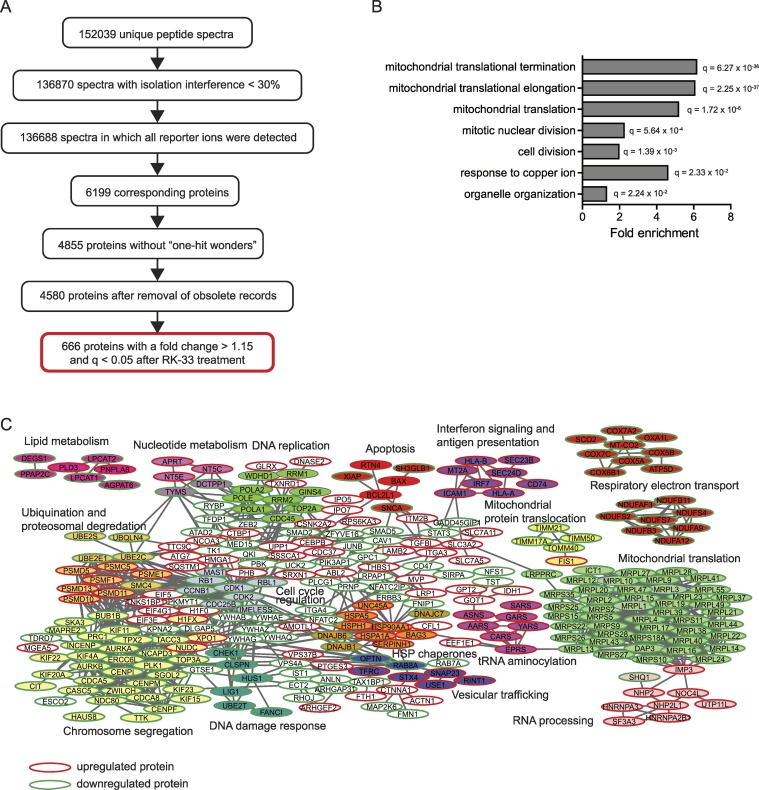
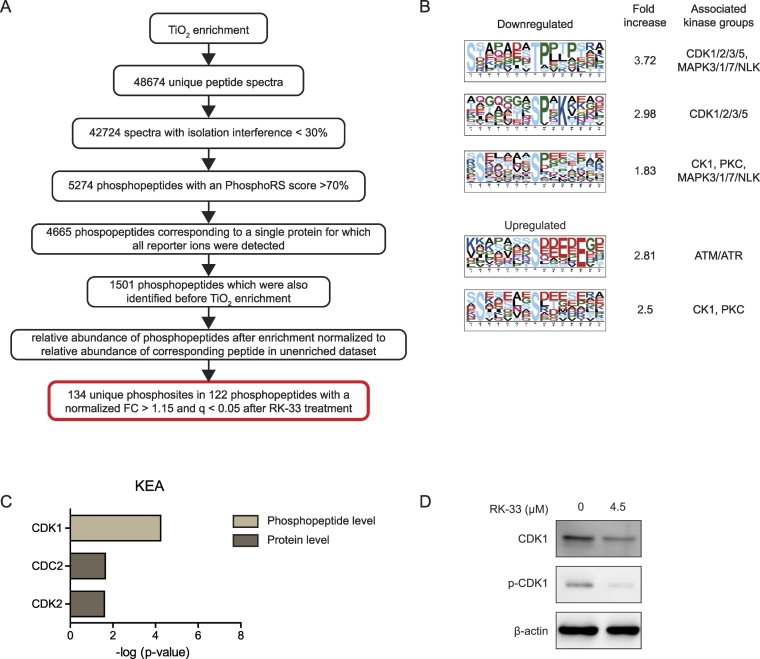
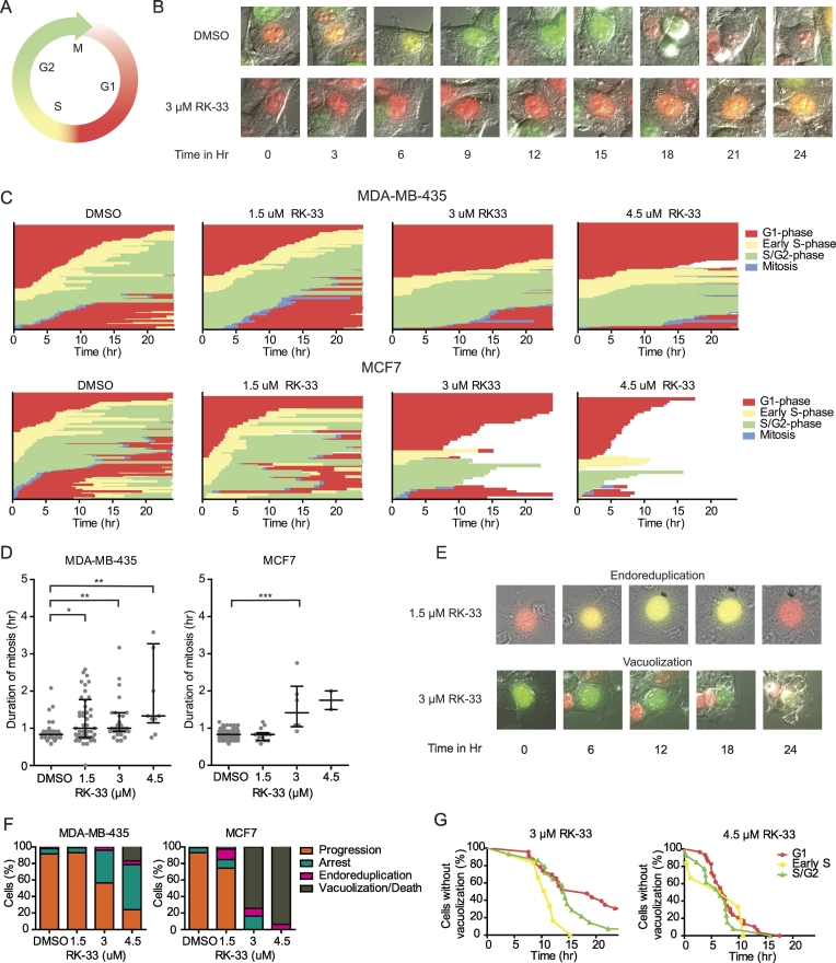
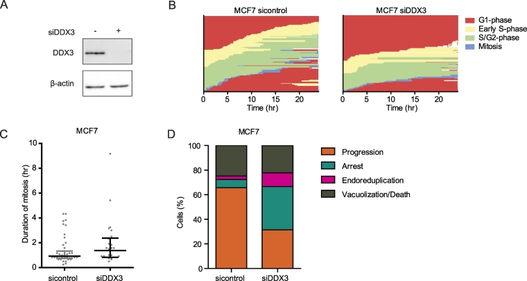
DDX3 is an RNA helicase with oncogenic properties. The small molecule inhibitor RK-33 is designed to fit into the ATP binding cleft of DDX3 and hereby block its activity. RK-33 has shown potent activity in preclinical cancer models. However, the mechanism behind the antineoplastic activity of RK-33 remains largely unknown. In this study we used a dual phosphoproteomic and single cell tracking approach to evaluate the effect of RK-33 on cancer cells. MDA-MB-435 cells were treated for 24 hours with RK-33 or vehicle control. Changes in phosphopeptide abundance were analyzed with quantitative mass spectrometry using isobaric mass tags (Tandem Mass Tags). At the proteome level we mainly observed changes in mitochondrial translation, cell division pathways and proteins related to cell cycle progression. Analysis of the phosphoproteome indicated decreased CDK1 activity after RK-33 treatment. To further evaluate the effect of DDX3 inhibition on cell cycle progression over time, we performed timelapse microscopy of Fluorescent Ubiquitin Cell Cycle Indicators labeled cells after RK-33 or siDDX3 exposure. Single cell tracking indicated that DDX3 inhibition resulted in a global delay in cell cycle progression in interphase and mitosis. In addition, we observed an increase in endoreduplication. Overall, we conclude that DDX3 inhibition affects cells in all phases and causes a global cell cycle progression delay.


Figures

Figure 1
Figure 1
The proteomic landscape after DDX3 inhibition with RK-33. A. Flowchart showing bioinformatical processing and analysis of peptide spectra identified by quantitative proteomics after 24 hours DMSO or 4.5 μM RK-33 exposure in MDA-MB-435 cells. B. Bar graph showing significantly enriched GOterms identified by gene-set enrichment analysis among proteins significantly altered after DDX3 inhibition with RK-33 treatment. Q-values are calculated are Benjamini and adjusted p-values generated by hypergeometric enrichment test. C. STRING network analysis showing clusters of interrelated proteins among the significantly altered proteins after RK-33 treatment. Each circle corresponds to a protein and is labeled with the gene symbol. The color fills indicate groups of proteins with a particular function (see labels). Direction of the fold change is indicated by the border color for each protein: green indicates downregulation, red indicates upregulation.
Figure 2
Figure 2
Changes in the phosphoproteome after RK-33 treatment indicate altered CDK1 activity. A. Flowchart showing bioinformatical processing and analysis of phospopeptide spectra identified by quantitative phospoproteomics after 24 hours DMSO or 4.5 μM RK-33 exposure in MDA-MB-435 cells. B. Motif-x analysis showing enriched motifs among the significantly up- or downregulated phosphopeptides and their associated kinase groups in the Networkin database. C. Bar graphs showing kinase enrichment analysis (KEA) among kinases known to phosphorylate the phosphopeptides and phosphoproteins significantly altered after RK-33 treatment. Benjamini and Hochberg corrected p-values of hypergeometric enrichment tests. D. Immunoblot showing a decrease in CDK1 and pCDK1 expression after 12 hours 4.5 μM RK-33 exposure in MDA-MB-435 cells.
Figure 3
Figure 3
Single cell tracking shows RK-33 causes a delay in all cell cycle phases. A. Schematic overview of cell cycle progression with fluorescent changes of the FUCCI constructs mCherry-hCdt1 and mVenus-hGeminin. B. Example merged DIC and fluorescent images showing the color change in a single DMSO and RK-33 treated MDA-MB-435-FUCCI cell over 24 hours. C. Graphs showing delayed cell cycle progression in single FUCCI labeled MDA-MB-435 and MCF7 cells after RK-33 treatment over 24 hours. Each line on the y-axis represents a single cell. D. Dot plot showing the median duration of mitosis after RK-33 treatment in FUCCI labeled MDA-MB-435 and MCF7 cells. Graphs represent median with interquartile range. P-values were calculated by a Mann–Whitney U Test. * P < .05, ** P < .01, *** P < .001. E. Example of endoreduplication and vacuolization occurring after RK-33 treatment in FUCCI-labeled MCF7 cells. F. Bar graphs showing the cell fate of RK-33 treated, FUCCI-labeled MCF7 and MDA-MB-435 cells. G. Kaplan Meijer plots showing the time to vacuolization or cell death for each phase during which the cell (MCF7) was first exposed to RK-33.
Figure 4
Figure 4
DDX3 knockdown results in a global cell cycle delay and endoreduplication. A. Immunoblot showing DDX3 expression 48 hours after siControl or siDDX3 transfection in MCF7 cells. B. Graphs showing delayed cell cycle progression in single MCF7-FUCCI cells 48 hours to 72 hours after siControl or siDDX3 transfection. Each line on the y-axis represents a single cell. C. Dot plot showing the median duration of mitosis after siDDX3 transfection in FUCCI labeled MCF7 cells. Graphs represent median with interquartile range. P-values were calculated by a Mann–Whitney U Test. D. Bar graphs showing the cell fate of MCF7-FUCCI cells 48 to 72 hours after siDDX3 transfection.

Similar articles

Cited by

References

    1. Linder P, Fuller-Pace FV. Looking back on the birth of DEAD-box RNA helicases. Biochim Biophys Acta. 2013;1829(8):750–755. - PubMed
    1. Botlagunta M, Vesuna F, Mironchik Y, Raman A, Lisok A, Winnard P, Jr., Mukadam S, Van Diest P, Chen JH, Farabaugh P. Oncogenic role of DDX3 in breast cancer biogenesis. Oncogene. 2008;27(28):3912–3922. - PMC - PubMed
    1. Heerma van Voss MR, Vesuna F, Trumpi K, Brilliant J, Berlinicke C, de Leng W, Kranenburg O, Offerhaus GJ, Burger H, van der Wall E. Identification of the DEAD box RNA helicase DDX3 as a therapeutic target in colorectal cancer. Oncotarget. 2015;6(29):28312–28326. - PMC - PubMed
    1. Bol GM, Vesuna F, Xie M, Zeng J, Aziz K, Gandhi N, Levine A, Irving A, Korz D, Tantravedi S. Targeting DDX3 with a small molecule inhibitor for lung cancer therapy. EMBO Mol Med. 2015;7(5):648–669. - PMC - PubMed
    1. Xie M, Vesuna F, Tantravedi S, Bol GM, Heerma van Voss MR, Nugent K, Malek R, Gabrielson KL, Van Diest PJ, Tran PT. RK-33 radiosensitizes prostate cancer cells by blocking the RNA helicase DDX3. Cancer Res. 2016 - PMC - PubMed