

A subjective and objective comparison of tissue contrast and imaging artifacts present in routine spin echoes and in iterative decomposition of asymmetric spin echoes for soft tissue neck MRI
- PMID: 29685536
- PMCID: PMC7453706
- DOI: 10.1016/j.ejrad.2018.03.016
A subjective and objective comparison of tissue contrast and imaging artifacts present in routine spin echoes and in iterative decomposition of asymmetric spin echoes for soft tissue neck MRI
Abstract
Objective: FSE sequences play key roles in neck MRI despite the susceptibility issues in neck region. Iterative decomposition of asymmetric echoes (IDEAL, GE) is a promising method that separates fat and water images resulting in high SNR and improved fat suppression. We tested how neck tissue contrasts, image artifacts and fat separation as opposed to fat suppression in terms of image quality compare between routine and IDEAL FSE.
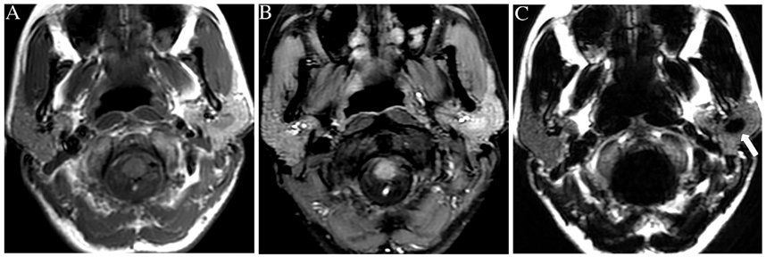
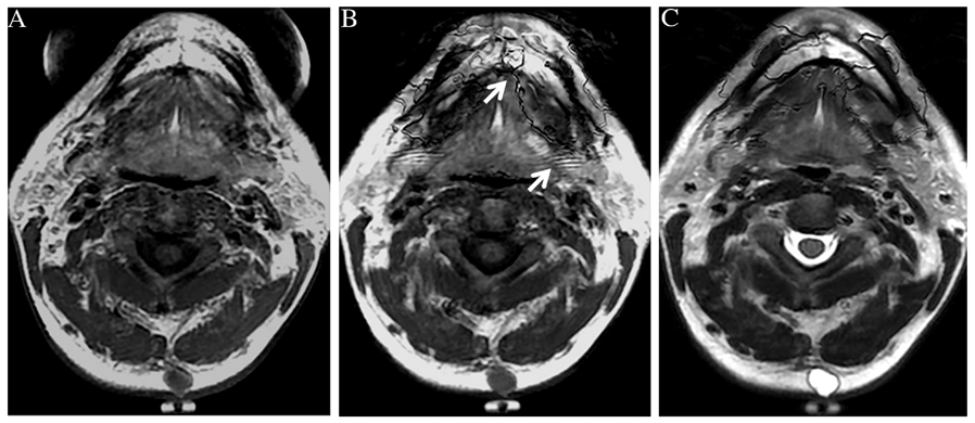
Methods: IDEAL based and routine T1 and T2-weighted FSE sequences were applied for neck MRI at 1.5T and 3T. Overall image quality including fat suppression, tissue contrast, image artifacts and lesion conspicuity were subjectively assessed for 20 patients clinically indicated for neck MRI. Quantitative tissue contrast estimates from parotid area were compared between IDEAL and routine FSE for 7 patients. Four patients with oncocytoma were also reviewed to assess benefits of separately reconstructed fat specific image sets.
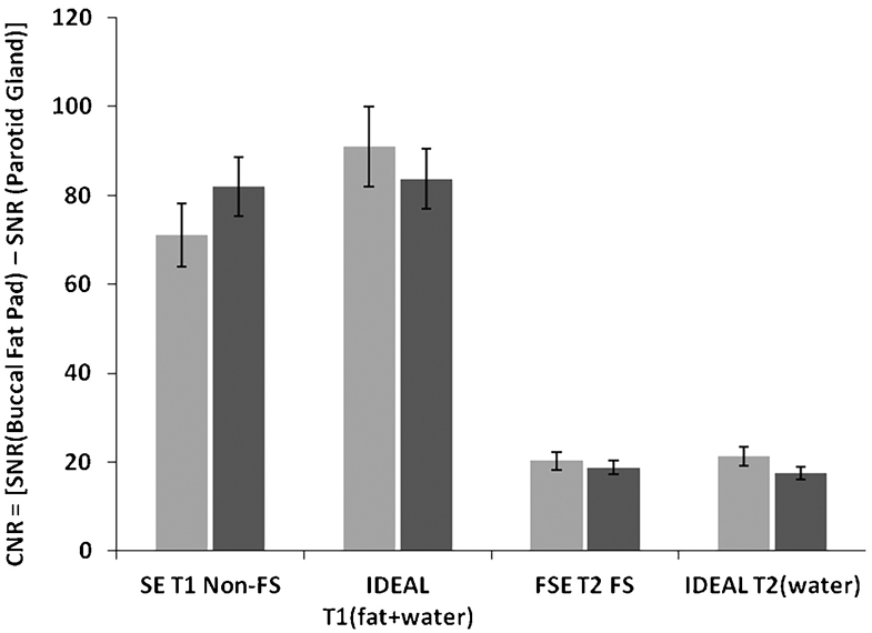
Results: Subjective tissue contrast and overall image quality including image sharpness, fat suppression and image artifacts were superior for IDEAL sequences. For oncocytoma fat specific IDEAL images provided additional information. Objective CNR estimates from a central slice were equivalent for IDEAL and routine FSE at both field strengths.
Conclusions: We demonstrated that high SNR inherent in IDEAL FSE consistently translates into high tissue contrast with image quality advantages in neck anatomy where large susceptibility variation and physiological motions reduce image quality for conventional FSE T1 and T2. However, the objective contrast estimates for parotid gland at isocenter were statistically equivalent for IDEAL and conventional FSE perhaps because at or near isocenter routine FSE works well. Additionally, fat specific IDEAL image sets add to diagnostic specificity for fat deficient lesions.
Keywords: Fat suppression; IDEAL; Image quality; Soft tissue neck; Susceptibility; Tissue contrast.
Copyright © 2018 Elsevier B.V. All rights reserved.
Conflict of interest statement
Figures
 at 3T,
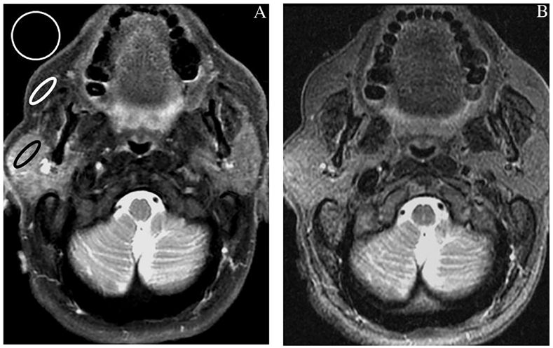
  at 1.5T. The mean CNR values for buccal fat/parotid are plotted from IDEAL and non-IDEAL FSE, ROI placement is shown in Figure 1.
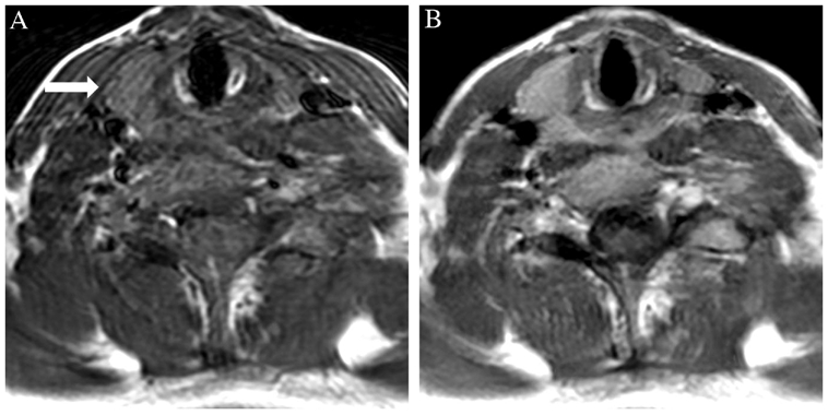
References
-
- Kitagawa Y, Ishii Y, Kawamura Y et al., Usefulness of fat-suppression magnetic resonance imaging for oral and maxillofacial lesions. Int. J. Oral and Maxillofac. Surg 25 (1996) 457–62. - PubMed
-
- Bydder GM, Pennock JM, Steiner RE et al., The short TI inversion recovery sequence—an approach to MR imaging of the abdomen. Magn. Reson. Imaging 3 (1985) 251–254. - PubMed
-
- Sadick M, Sadick H, Hörmann K et al., Diagnostic evaluation of magnetic resonance imaging with turbo inversion recovery sequence in head and neck tumors. Eur. Arch. Otorhinolaryngol 262 (2005) 634–39. - PubMed
-
- Nakatsu M, Hatabu H, Itoh H et al., Comparison of short inversion time inversion recovery (STIR) and fat-saturated (chemsat) techniques for background fat intensity suppression in cervical and thoracic MR imaging. JMRI J. Magn. Reson. Imaging 11 (2000) 56–60. - PubMed
-
- Sarkar SN, Komoroski R, NMR Imaging of Morphology, Defects, and Composition of Tire Composites and Model Elastomer Blends. Macromolecules 25 (1992) 1420–26.
Publication types
MeSH terms
Grants and funding
LinkOut - more resources
Full Text Sources
Other Literature Sources
