. 2018 May 8;115(19):4969-4974.
doi: 10.1073/pnas.1802279115. Epub 2018 Apr 23.

Prevention of hepatocellular carcinoma by targeting MYCN-positive liver cancer stem cells with acyclic retinoid

Affiliations

Prevention of hepatocellular carcinoma by targeting MYCN-positive liver cancer stem cells with acyclic retinoid

Xian-Yang Qin et al. Proc Natl Acad Sci U S A. .

Abstract

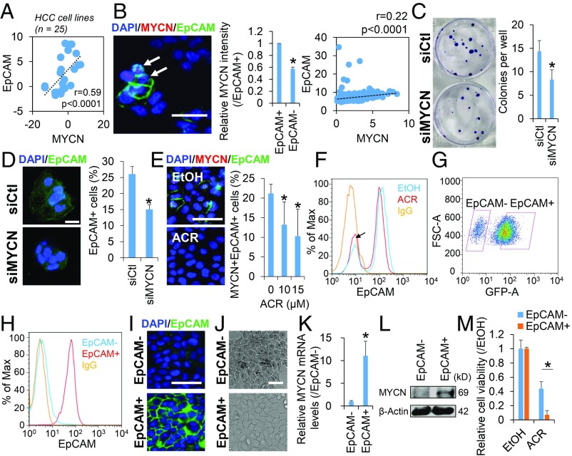
Hepatocellular carcinoma (HCC) is a highly lethal cancer that has a high rate of recurrence, in part because of cancer stem cell (CSC)-dependent field cancerization. Acyclic retinoid (ACR) is a synthetic vitamin A-like compound capable of preventing the recurrence of HCC. Here, we performed a genome-wide transcriptome screen and showed that ACR selectively suppressed the expression of MYCN, a member of the MYC family of basic helix-loop-helix-zipper transcription factors, in HCC cell cultures, animal models, and liver biopsies obtained from HCC patients. MYCN expression in human HCC was correlated positively with both CSC and Wnt/β-catenin signaling markers but negatively with mature hepatocyte markers. Functional analysis showed repressed cell-cycle progression, proliferation, and colony formation, activated caspase-8, and induced cell death in HCC cells following silencing of MYCN expression. High-content single-cell imaging analysis and flow cytometric analysis identified a MYCN+ CSC subpopulation in the heterogeneous HCC cell cultures and showed that these cells were selectively killed by ACR. Particularly, EpCAM+ cells isolated using a cell-sorting system showed increased MYCN expression and sensitivity to ACR compared with EpCAM- cells. In a long-term (>10 y) follow-up study of 102 patients with HCC, MYCN was expressed at higher levels in the HCC tumor region than in nontumor regions, and there was a positive correlation between MYCN expression and recurrence of de novo HCC but not metastatic HCC after curative treatment. In summary, these results suggest that MYCN serves as a prognostic biomarker and therapeutic target of ACR for liver CSCs in de novo HCC.

Keywords: MYCN; acyclic retinoid; cancer stem cell; hepatocellular carcinoma; transcriptome.


Conflict of interest statement

The authors declare no conflict of interest.

Figures

Fig. 1.
Fig. 1.
Identification of MYCN as a molecular target of ACR. (A) Heatmap of the top 10 up-regulated and down-regulated genes that were differentially expressed in JHH7 cells and Hc cells 1 and 4 h after starting treatment with 1 μM atRA (control, Ctl) or 10 μM ACR as assessed by CAGE analysis. The genes were ranked by the fold change when ACR treatment was compared with low-dose atRA treatment. (B, Upper) Immunofluorescence staining for MYCN in JHH7 cell cultures treated with 0.05% ethanol (EtOH, vehicle) or 10 μM ACR for 24 h (n = 3). (Scale bar, 50 μm.) (Lower Left) Percentages of MYCN+ cells among the total number of JHH7 cell cultures counted. (Lower Right) The relative fluorescent intensity of MYCN protein vs. EtOH. (C) c-MYC (Upper) and MYCN (Lower) gene expression as assessed by CAGE analysis. TPM, tags per million mapped reads. (D) Chemical structures of ACR, its ester analogs, and atRA. (E and F) The effects of ACR and its ester analogs on cell viability 24 h after treatment (E) and MYCN gene expression levels 4 h after treatment (10 μM each) (F) in JHH7 cell cultures (n = 3). (G and H) Schematic overview of experimental procedures and time-dependent incidence of liver tumor (G) and MYCN gene expression in the livers (H) of a diet-induced mouse model of NASH (n = 6–10 mice per group). Ath+HF, atherogenic and high fat diet; LF, low fat diet. Purple circles indicate the mice suffering from liver tumor. The data are presented as the mean ± SD; *P < 0.05, Student’s t test. n.s., not significant.
Fig. 2.
Fig. 2.
Loss-of-function analysis of MYCN in JHH7 cells. Cells in the G1 and G2 phases were obtained through flow cytometric cell sorting of Hoechst 33342-stained JHH7 cells. (A and B) Cyclin B (A) and MYCN (B) gene expression in sorted cells. (C) MYCN gene expression in JHH7 cells transfected with either control siRNA (siCtl) or siRNA to MYCN (siMYCN, three target-specific siRNAs targeting human MYCN) for 72 h. (DF)The cells were reseeded in 96-well plates, and 72 h later the cell-cycle stages (D) and percentages of Ki67+ (E) or clCasp3+ (F) cells in the JHH7 cell cultures were examined using imaging analysis. (G and H) The time-dependent effect of MYCN knockdown on cell viability (G) and Casp8 activity (H) in JHH7 cell cultures. (I and J) Casp8 activity (I) and cell viability (J) in JHH7 cell cultures that were treated with either 0.05% EtOH or 10 μM ACR for 24 h in the absence (−) or presence (+) of 20 μM z-IETD-FMK, a Casp8 inhibitor. (Scale bars, 50 μm.) The data are presented as the mean ± SD (n = 3–4). *P < 0.05, Student’s t test. siCtl, control siRNA.
Fig. 3.
Fig. 3.
The MYCN-positive CSC subpopulation of JHH7 cells is selectively targeted by ACR. (A) Correlation between gene expression of MYCN and EpCAM in a total of 25 HCC cell lines in the CCLE database. The data are presented as a robust multiarray average (RMA). (B, Left) Immunofluorescence triple staining of DAPI (blue), MYCN (red), and a liver CSC marker EpCAM (green) in JHH7 cell cultures. (Scale bar, 20 μm.) (Center) MYCN intensity in EpCAM+ and EpCAM cells. (Right) The correlation between the intensities of MYCN and EpCAM in individual cells was assessed using the Pearson’s correlation coefficient. (C and D) The inhibitory effect of MYCN knockdown on the colony-forming ability of JHH7 cells was evaluated in a limiting dilution assay (C) and by the percentage of EpCAM+ cells in JHH7 cell cultures (D). (Scale bar, 20 μm.) (E) A dose-dependent inhibitory effect of ACR treatment for 24 h on the percentage of MYCN+EpCAM+ cells in JHH7 cell cultures. (Scale bar, 50 μm.) (F) Flow cytometric analysis of EpCAM expression in JHH7 cells treated with 15 μM ACR for 24 h. Dead cells were excluded by propidium iodide (PI) staining. The arrow indicates the increased presence of EpCAM cells following ACR treatment. (G) EpCAM+ and EpCAM cells were obtained through flow cytometric cell sorting of EpCAM-Alexa 488–stained JHH7 cells. (HJ) The sorted cells were reanalyzed in flow cytometric analysis (H) and were characterized in immunofluorescence (I) and brightfield microscopic (J) analysis. (Scale bars, 50 μm.) (K and L) Gene (K) and protein (L) expression of MYCN. (M) Effect of ACR treatment at 10 μM for 48 h on the cell viability in the sorted cells. The data are presented as the mean ± SD (n = 3 or 4). *P < 0.05, Student’s t test.
Fig. 4.
Fig. 4.
MYCN is a prognostic factor for the recurrence of de novo HCC. (A, Upper) Schematic overview of a clinical study that enrolled 12 HCC patients who received 8 wk of ACR administration (300 mg/d or 600 mg/d) after definitive treatment (n = 6 per group). (Lower) The ratio of HCC patients who showed decreased MYCN expression in their liver biopsies (<0.5-fold) after ACR treatment. (B) Representative images of H&E staining and immunohistochemical staining of MYCN in liver sections of nontumor adjacent (N) and HCC (T) tissues. (Scale bar, 100 μm.) (C) MYCN gene expression in normal liver tissues collected at a distance from a liver metastasis of colon cancer (Ctl, n = 5) and matched nontumor adjacent (N, n = 50) and HCC (T, n = 50) liver tissues as assessed by CAGE analysis in a European cohort. (D and E) MYCN gene-expression levels in surgical matched nontumor adjacent (N) and HCC (T) liver tissues in all the enrolled HCC patients (n = 102) (D) or in subpopulations of patients without HCC recurrence (non-Rec, n = 24) or with HCC recurrence (Rec, n = 78) (E) during a long-term follow-up (>10 y) after curative treatment in a Japanese cohort. (F) Prognostic significance of MYCN expression in human HCC assessed using the Kaplan–Meier method and log-rank test in subpopulations of patients without intrahepatic metastasis (n = 74) at the time of curative treatment. Low MYCN, HCC patients with MYCN expression lower than the median expression value. High MYCN, HCC patients with MYCN expression equal to or higher than the median expression value. *P < 0.05, Student’s t test.

Comment in

Similar articles

Cited by

References

    1. Venook AP, Papandreou C, Furuse J, de Guevara LL. The incidence and epidemiology of hepatocellular carcinoma: A global and regional perspective. Oncologist. 2010;15:5–13. - PubMed
    1. Perz JF, Armstrong GL, Farrington LA, Hutin YJ, Bell BP. The contributions of hepatitis B virus and hepatitis C virus infections to cirrhosis and primary liver cancer worldwide. J Hepatol. 2006;45:529–538. - PubMed
    1. Yoshimoto S, et al. Obesity-induced gut microbial metabolite promotes liver cancer through senescence secretome. Nature. 2013;499:97–101, and erratum (2014) 506:396. - PubMed
    1. Kang TW, et al. Senescence surveillance of pre-malignant hepatocytes limits liver cancer development. Nature. 2011;479:547–551. - PubMed
    1. Ma C, et al. NAFLD causes selective CD4(+) T lymphocyte loss and promotes hepatocarcinogenesis. Nature. 2016;531:253–257. - PMC - PubMed

Publication types

MeSH terms

Substances

LinkOut - more resources