. 2018 May 29;115(22):E5213-E5222.
doi: 10.1073/pnas.1722335115. Epub 2018 Apr 23.

MYB72-dependent coumarin exudation shapes root microbiome assembly to promote plant health

Affiliations

MYB72-dependent coumarin exudation shapes root microbiome assembly to promote plant health

Ioannis A Stringlis et al. Proc Natl Acad Sci U S A. .

Abstract

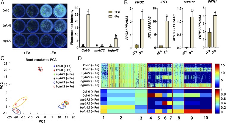
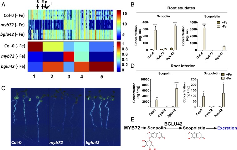
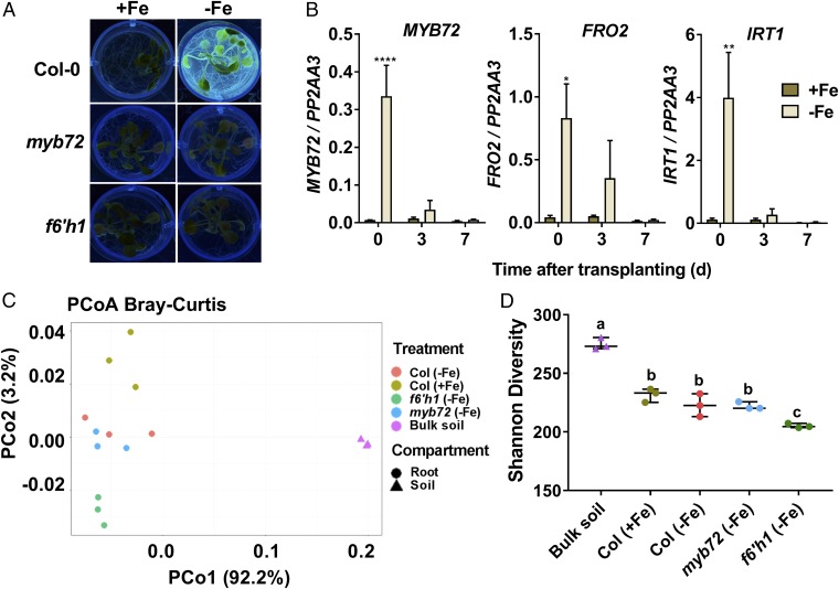
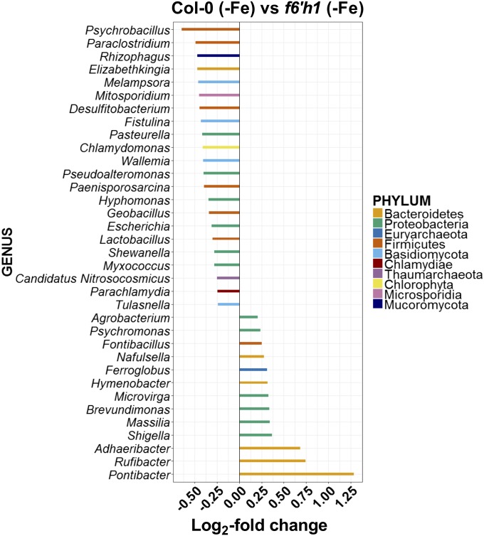
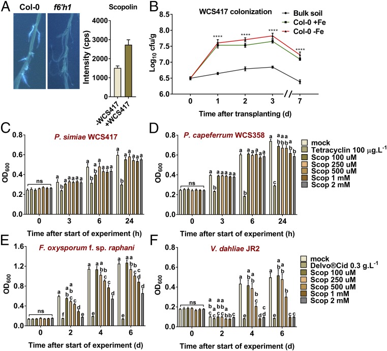
Plant roots nurture a tremendous diversity of microbes via exudation of photosynthetically fixed carbon sources. In turn, probiotic members of the root microbiome promote plant growth and protect the host plant against pathogens and pests. In the Arabidopsis thaliana-Pseudomonas simiae WCS417 model system the root-specific transcription factor MYB72 and the MYB72-controlled β-glucosidase BGLU42 emerged as important regulators of beneficial rhizobacteria-induced systemic resistance (ISR) and iron-uptake responses. MYB72 regulates the biosynthesis of iron-mobilizing fluorescent phenolic compounds, after which BGLU42 activity is required for their excretion into the rhizosphere. Metabolite fingerprinting revealed the antimicrobial coumarin scopoletin as a dominant metabolite that is produced in the roots and excreted into the rhizosphere in a MYB72- and BGLU42-dependent manner. Shotgun-metagenome sequencing of root-associated microbiota of Col-0, myb72, and the scopoletin biosynthesis mutant f6'h1 showed that scopoletin selectively impacts the assembly of the microbial community in the rhizosphere. We show that scopoletin selectively inhibits the soil-borne fungal pathogens Fusarium oxysporum and Verticillium dahliae, while the growth-promoting and ISR-inducing rhizobacteria P. simiae WCS417 and Pseudomonas capeferrum WCS358 are highly tolerant of the antimicrobial effect of scopoletin. Collectively, our results demonstrate a role for coumarins in microbiome assembly and point to a scenario in which plants and probiotic rhizobacteria join forces to trigger MYB72/BGLU42-dependent scopolin production and scopoletin excretion, resulting in improved niche establishment for the microbial partner and growth and immunity benefits for the host plant.

Keywords: coumarin; induced systemic resistance; iron-deficiency response; microbiome assembly; root metabolome.


Conflict of interest statement

The authors declare no conflict of interest.

Figures

Fig. 1.
Fig. 1.
Metabolite fingerprinting of root exudates of Col-0, myb72, and bglu42 plants grown under iron-sufficient and iron-starvation conditions. (A) Accumulation and secretion of fluorescent phenolic compounds of 20-d-old Col-0, myb72, and bglu42 plants grown in Hoagland medium with (+Fe) or without (−Fe) iron. Visualization of fluorescence was achieved under UV light (365 nm). Fluorescence intensity was quantified in a 96-well microplate reader (excitation: 360 nm; emission: 528 nm) after seedlings were removed from the growth medium. Different letters indicate significant differences (P < 0.05, Tukey’s test, two-way ANOVA). (B) Gene-expression profiles of the iron-deficiency marker genes FRO2, IRT1, MYB72, and F6′H1 in roots of 20-d-old Col-0 plants grown in Hoagland medium with (+Fe) or without (−Fe) iron, quantified by qRT-PCR. Transcript levels were normalized to that of the reference gene PP2AA3 (At1g13320). Data are the means of three biological replicates. Error bars represent the SEM. Asterisks indicate significant differences between treatments: **P < 0.001, *P < 0.05, Student’s t test. (C) PCA plot of root exudates of 20-d-old Col-0 (encircled in yellow), myb72 (encircled in red), and bglu42 (encircled in blue) plants grown in Hoagland medium with (+Fe) or without (−Fe) iron. Data were obtained by metabolite fingerprinting (UPLC-ESI-TOF-MS analysis in positive and negative ionization mode), and the PCA represents the data subset after the filtering. PC1, principal component 1; PC2, principal component 2. (D) 1D-SOM clustering and prototype assignment of 722 high-quality metabolite features (FDR <0.001) derived from the positive as well as the negative ionization mode of the metabolite fingerprinting analysis. The number of features assigned to one prototype determines its width. The raw intensity of individual features (Upper) and prototypes (Lower) mostly affected by iron deficiency are highlighted in the red boxes. Heatmaps correspond to the intensity of each individual feature (Upper) and the average intensity of each cluster/prototype (Lower). The color key shows the range of signal intensities. Three biological replicates per treatment were used for analysis.
Fig. 2.
Fig. 2.
MYB72- and BGLU42-dependent metabolites in root exudates and roots of Col-0, myb72, and bglu42 plants. (A) 1D-SOM clustering and prototype assignment of 311 high-quality metabolite features (FDR <0.001) in root exudates of iron-starved Col-0, myb72, and bglu42 plants. The number of features assigned to one prototype determines its width. Heatmaps correspond to the intensity of each individual feature (Upper) and the average intensity of each cluster/prototype (Lower). The color key shows the range of signal intensities. Arrows indicate the position of scopoletin (S), esculin (E), esculetin (e), and isofraxidin (I). For details on selected metabolite features, see Fig. S2 and Table S1. (B) HPLC-DAD quantification of scopolin and scopoletin in root exudates of Col-0, myb72, and bglu42 plants grown under iron-sufficient (+Fe) and iron-starved (−Fe) conditions. The data are the means of three replicates of the pooled root exudates of 50–60 plants per replicate. Error bars represent the SEM. Asterisks indicate significant differences between the iron conditions within a genotype: ****P < 0.0001, **P < 0.01, *P < 0.05, two-way ANOVA, Sidak’s test. (C) Photographs of iron-starved Col-0, myb72, and bglu42 plants grown in 12-well plates with liquid Hoagland medium without iron. Visualization of fluorescent phenolic compounds was achieved under UV light (365 nm). (D) HPLC-DAD quantification of scopolin and scopoletin in the roots of Col-0, myb72, and bglu42 plants grown under iron-sufficient and iron-starved conditions. The data are the means of three replicates of the pooled root extracts of 130 plants per replicate. Error bars represent SE. Asterisks indicate significant differences between the iron conditions within a genotype: ****P < 0.0001, **P < 0.01, *P < 0.05, two-way ANOVA, Sidak’s test. (E) Schematic representation of the role of MYB72 in the production of coumarin scopolin and the activity of BGLU42 in the deglycosylation of scopolin and the subsequent production of the aglycone scopoletin before its excretion into the rhizosphere. The presented molecules were created using the website https://www.emolecules.com.
Fig. 3.
Fig. 3.
Metagenome analysis of bulk soil and root-associated microbiomes of Col-0, myb72, and f6′h1 plants. (A) Excretion of fluorescent phenolic compounds by 26-d-old Col-0, myb72, and f6′h1 plants grown in Hoagland medium with (+Fe) or without (−Fe) iron. Visualization of fluorescence was achieved under UV light (365 nm). (B) Gene-expression profiles of MYB72 and the iron-deficiency marker genes FRO2 and IRT1 in roots of Col-0 plants pregrown for 14 d in Hoagland medium with (+Fe) or without (−Fe) iron and transplanted on day 0 to limed Reijerscamp soil. Gene expression was quantified by qRT-PCR. Transcript levels were normalized to that of reference gene PP2AA3 (At1g13320). Data are means of three biological replicates. Error bars represent the SEM. Asterisks indicate significant differences between treatments: ****P < 0.0001, **P < 0.01, *P < 0.05, two-way ANOVA, Sidak’s test. (C) PCoA using Bray–Curtis metrics displays the dissimilarity of microbial communities in soil (triangles) and root samples (circles) and of Col-0 plants pregrown under iron-sufficient (+Fe) conditions and Col-0, myb72, and f6′h1 plants pregrown under iron-starved conditions (−Fe). (D) Shannon diversity (effective number of species) displaying the within-sample diversity of different treatments. Horizontal bars correspond to the median and interquartile range of values. Different letters indicate significant differences: P < 0.05, one-way ANOVA, Tukey’s test.
Fig. 4.
Fig. 4.
Differential abundance of microbial genera on Arabidopsis roots with different scopoletin exudation patterns. Differentially abundant bacterial and fungal genera in root samples of Col-0 (−Fe) and f6′h1 (−Fe) plants as determined using DESeq2. Comparisons were performed at the genus level using an FDR <0.05 to select for significance. In all graphs, negative log2 fold-change values relate to genera that are significantly enriched in iron-starved Col-0 (−Fe) root samples in comparison with the contrasting genotype/treatment combination. Different bar colors represent different phyla as shown in the key.
Fig. 5.
Fig. 5.
Effect of scopoletin on P. simiae WCS417 colonization and on the growth of selected soil-inhabiting microbes. (A, Left) Visualization of fluorescent phenolic compounds produced by roots of iron-sufficient Col-0 and scopoletin biosynthesis mutant f6′h1 plants in response to colonization by P. simiae WCS417. Visualization of fluorescent phenolic compounds was achieved under UV light (365 nm). Photographs were taken from roots of 20-d-old in vitro-grown Arabidopsis plants 7 d after colonization by the rhizobacteria. (Right) Bars show signal intensity in counts per second of the coumarin scopolin (detected as [M+H]+) in Col-0 roots 2 d after colonization by P. simiae WCS417. Shown data are the means (± SEM) of three biological replicates (see also Fig. S5). (B) Number of P. simiae WCS417 bacteria recovered from rhizospheres of Col-0 plants grown in limed Reijerscamp soil that was amended with 105 cfu⋅g−1 WCS417 bacteria. Root colonization was determined at the indicated days after the seedlings were transplanted from iron-sufficient (+Fe) or iron-starved (−Fe) Hoagland growth medium into the WCS417-amended Reijerscamp soil. Values for each time point were calculated from five rhizosphere or bulk soil samples. Asterisks indicate significant differences between bulk soil and colonized plants: ****P < 0.0001, two-way ANOVA, Tukey’s test. (CF) Graphs showing growth (OD600) of P. simiae WCS417 (C), P. capeferrum WCS358 (D), F. oxysporum f.sp. raphani (E), and V. dahliae JR2 (F) in medium containing the indicated concentrations of scopoletin (Scop). Tetracycline and Delvocid were used as positive controls for bacteria and fungi, respectively. Growth measurements were performed over a period of 24 h (bacteria) or 6 d (fungi). The data shown are the means of 8–10 replicates. Error bars represent the SE of the mean. Different letters represent significant differences between treatments: P < 0.05, two-way ANOVA, Tukey’s test.

Comment in

  • Root-exuded coumarin shapes the root microbiome.
    Lundberg DS, Teixeira PJPL. Lundberg DS, et al. Proc Natl Acad Sci U S A. 2018 May 29;115(22):5629-5631. doi: 10.1073/pnas.1805944115. Epub 2018 May 15. Proc Natl Acad Sci U S A. 2018. PMID: 29764997 Free PMC article. No abstract available.

References

    1. Bais HP, Weir TL, Perry LG, Gilroy S, Vivanco JM. The role of root exudates in rhizosphere interactions with plants and other organisms. Annu Rev Plant Biol. 2006;57:233–266. - PubMed
    1. Berendsen RL, Pieterse CMJ, Bakker PAHM. The rhizosphere microbiome and plant health. Trends Plant Sci. 2012;17:478–486. - PubMed
    1. Mendes R, et al. Deciphering the rhizosphere microbiome for disease-suppressive bacteria. Science. 2011;332:1097–1100. - PubMed
    1. Bulgarelli D, Schlaeppi K, Spaepen S, Ver Loren van Themaat E, Schulze-Lefert P. Structure and functions of the bacterial microbiota of plants. Annu Rev Plant Biol. 2013;64:807–838. - PubMed
    1. Hacquard S, Spaepen S, Garrido-Oter R, Schulze-Lefert P. Interplay between innate immunity and the plant microbiota. Annu Rev Phytopathol. 2017;55:565–589. - PubMed

Publication types