

Nonviral gene editing via CRISPR/Cas9 delivery by membrane-disruptive and endosomolytic helical polypeptide
- PMID: 29686087
- PMCID: PMC5948953
- DOI: 10.1073/pnas.1712963115
Nonviral gene editing via CRISPR/Cas9 delivery by membrane-disruptive and endosomolytic helical polypeptide
Abstract
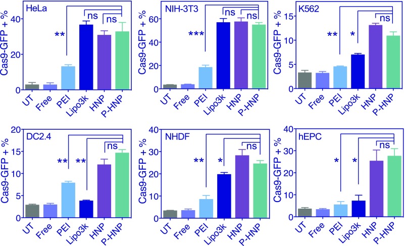
Effective and safe delivery of the CRISPR/Cas9 gene-editing elements remains a challenge. Here we report the development of PEGylated nanoparticles (named P-HNPs) based on the cationic α-helical polypeptide poly(γ-4-((2-(piperidin-1-yl)ethyl)aminomethyl)benzyl-l-glutamate) for the delivery of Cas9 expression plasmid and sgRNA to various cell types and gene-editing scenarios. The cell-penetrating α-helical polypeptide enhanced cellular uptake and promoted escape of pCas9 and/or sgRNA from the endosome and transport into the nucleus. The colloidally stable P-HNPs achieved a Cas9 transfection efficiency up to 60% and sgRNA uptake efficiency of 67.4%, representing an improvement over existing polycation-based gene delivery systems. After performing single or multiplex gene editing with an efficiency up to 47.3% in vitro, we demonstrated that P-HNPs delivering Cas9 plasmid/sgRNA targeting the polo-like kinase 1 (Plk1) gene achieved 35% gene deletion in HeLa tumor tissue to reduce the Plk1 protein level by 66.7%, thereby suppressing the tumor growth by >71% and prolonging the animal survival rate to 60% within 60 days. Capable of delivering Cas9 plasmids to various cell types to achieve multiplex gene knock-out, gene knock-in, and gene activation in vitro and in vivo, the P-HNP system offers a versatile gene-editing platform for biological research and therapeutic applications.
Keywords: CRISPR/Cas9; cell-penetrating peptide; genome editing; helical polypeptide; nanomedicine.
Copyright © 2018 the Author(s). Published by PNAS.
Conflict of interest statement
The authors declare no conflict of interest.
Figures


Similar articles
-
Cationic Polymer-Mediated CRISPR/Cas9 Plasmid Delivery for Genome Editing.Macromol Rapid Commun. 2019 Mar;40(5):e1800068. doi: 10.1002/marc.201800068. Epub 2018 Apr 30. Macromol Rapid Commun. 2019. PMID: 29708298
-
Scaffold-mediated non-viral delivery platform for CRISPR/Cas9-based genome editing.Acta Biomater. 2019 May;90:60-70. doi: 10.1016/j.actbio.2019.04.020. Epub 2019 Apr 9. Acta Biomater. 2019. PMID: 30978509
-
Comparative analysis of lipid Nanoparticle-Mediated delivery of CRISPR-Cas9 RNP versus mRNA/sgRNA for gene editing in vitro and in vivo.Eur J Pharm Biopharm. 2024 Mar;196:114207. doi: 10.1016/j.ejpb.2024.114207. Epub 2024 Feb 6. Eur J Pharm Biopharm. 2024. PMID: 38325664
-
Nanoparticle-Based Delivery of CRISPR/Cas9 Genome-Editing Therapeutics.AAPS J. 2018 Oct 10;20(6):108. doi: 10.1208/s12248-018-0267-9. AAPS J. 2018. PMID: 30306365 Free PMC article. Review.
-
Delivery of CRISPR/Cas9 for therapeutic genome editing.J Gene Med. 2019 Jul;21(7):e3107. doi: 10.1002/jgm.3107. J Gene Med. 2019. PMID: 31237055 Review.
Cited by
-
Exploring nano-enabled CRISPR-Cas-powered strategies for efficient diagnostics and treatment of infectious diseases.J Nanostructure Chem. 2022;12(5):833-864. doi: 10.1007/s40097-022-00472-7. Epub 2022 Feb 14. J Nanostructure Chem. 2022. PMID: 35194511 Free PMC article. Review.
-
Gene Therapy for Cardiovascular Disease: Basic Research and Clinical Prospects.Front Cardiovasc Med. 2021 Nov 5;8:760140. doi: 10.3389/fcvm.2021.760140. eCollection 2021. Front Cardiovasc Med. 2021. PMID: 34805315 Free PMC article. Review.
-
Toward the Treatment of Inherited Diseases of the Retina Using CRISPR-Based Gene Editing.Front Med (Lausanne). 2021 Oct 1;8:698521. doi: 10.3389/fmed.2021.698521. eCollection 2021. Front Med (Lausanne). 2021. PMID: 34660621 Free PMC article. Review.
-
Amino Acids, Peptides, and Proteins: Implications for Nanotechnological Applications in Biosensing and Drug/Gene Delivery.Nanomaterials (Basel). 2021 Nov 8;11(11):3002. doi: 10.3390/nano11113002. Nanomaterials (Basel). 2021. PMID: 34835766 Free PMC article. Review.
-
Tissue Engineering and Regenerative Medicine: Achievements, Future, and Sustainability in Asia.Front Bioeng Biotechnol. 2020 Mar 24;8:83. doi: 10.3389/fbioe.2020.00083. eCollection 2020. Front Bioeng Biotechnol. 2020. PMID: 32266221 Free PMC article. Review.
References
-
- Wang HX, et al. CRISPR/Cas9-based genome editing for disease modeling and therapy: Challenges and opportunities for nonviral delivery. Chem Rev. 2017;117:9874–9906. - PubMed
-
- Wells DJ. Gene therapy progress and prospects: Electroporation and other physical methods. Gene Ther. 2004;11:1363–1369. - PubMed
Publication types
MeSH terms
Substances
Grants and funding
LinkOut - more resources
Full Text Sources
Other Literature Sources
Miscellaneous
