. 2018 Jun 11;200(13):e00690-17.
doi: 10.1128/JB.00690-17. Print 2018 Jul 1.

Domains within RbpA Serve Specific Functional Roles That Regulate the Expression of Distinct Mycobacterial Gene Subsets

Affiliations

Domains within RbpA Serve Specific Functional Roles That Regulate the Expression of Distinct Mycobacterial Gene Subsets

Jerome Prusa et al. J Bacteriol. .

Abstract

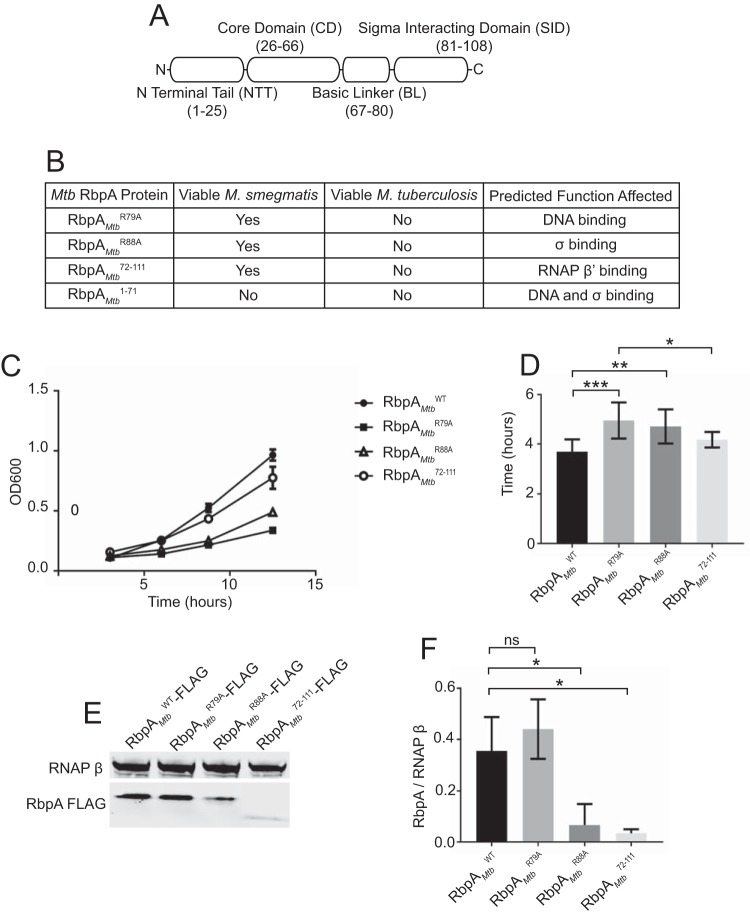
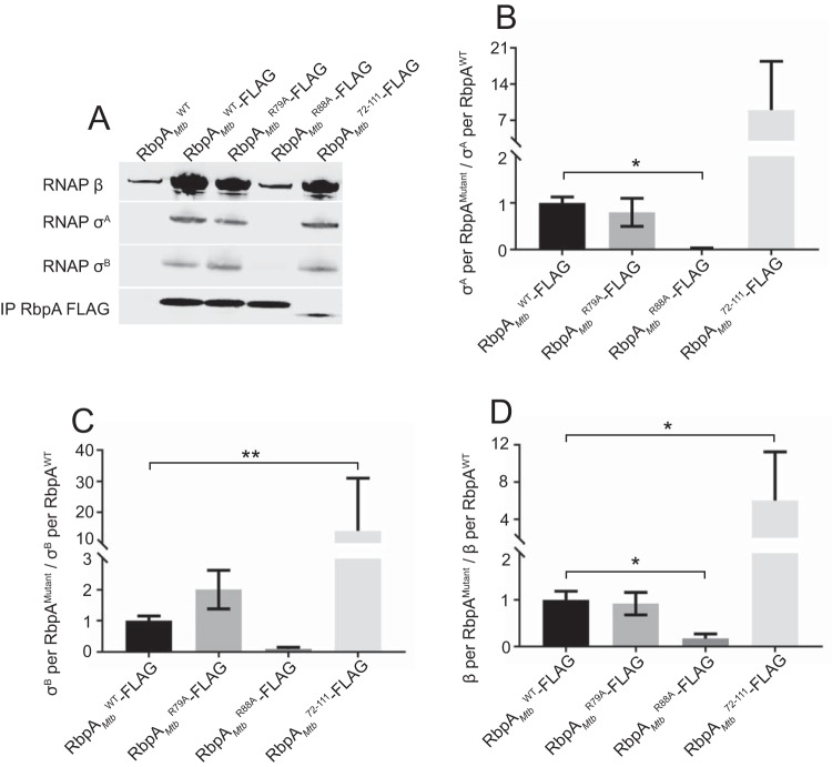
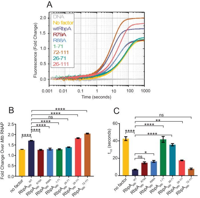
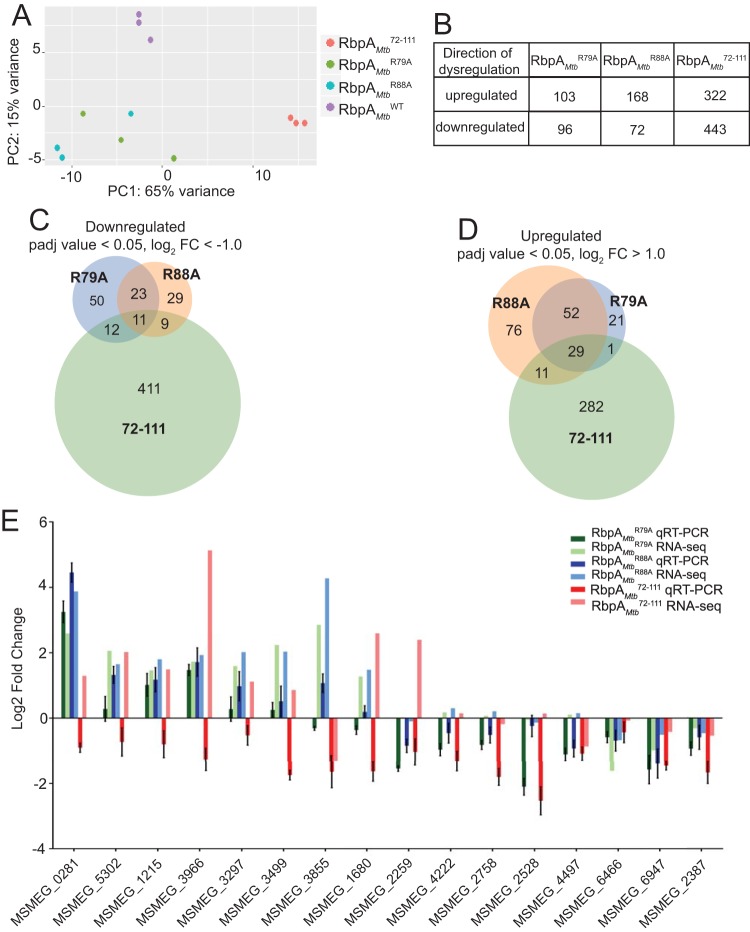
The RNA polymerase (RNAP) binding protein A (RbpA) contributes to the formation of stable RNAP-promoter open complexes (RPo) and is essential for viability in mycobacteria. Four domains have been identified in the RbpA protein, i.e., an N-terminal tail (NTT) that interacts with RNAP β' and σ subunits, a core domain (CD) that contacts the RNAP β' subunit, a basic linker (BL) that binds DNA, and a σ-interaction domain (SID) that binds group I and group II σ factors. Limited in vivo studies have been performed in mycobacteria, however, and how individual structural domains of RbpA contribute to RbpA function and mycobacterial gene expression remains mostly unknown. We investigated the roles of the RbpA structural domains in mycobacteria using a panel of rbpA mutants that target individual RbpA domains. The function of each RbpA domain was required for Mycobacterium tuberculosis viability and optimal growth in Mycobacterium smegmatis We determined that the RbpA SID is both necessary and sufficient for RbpA interaction with the RNAP, indicating that the primary functions of the NTT and CD are not solely association with the RNAP. We show that the RbpA BL and SID are required for RPo stabilization in vitro, while the NTT and CD antagonize this activity. Finally, RNA-sequencing analyses suggest that the NTT and CD broadly activate gene expression, whereas the BL and SID activate or repress gene expression in a gene-dependent manner for a subset of mycobacterial genes. Our findings highlight specific outcomes for the activities of the individual functional domains in RbpA.IMPORTANCEMycobacterium tuberculosis is the causative agent of tuberculosis and continues to be the most lethal infectious disease worldwide. Improved molecular understanding of the essential proteins involved in M. tuberculosis transcription, such as RbpA, could provide targets for much needed future therapeutic agents aimed at combatting this pathogen. In this study, we expand our understanding of RbpA by identifying the RbpA structural domains responsible for the interaction of RbpA with the RNAP and the effects of RbpA on transcription initiation and gene expression. These experiments expand our knowledge of RbpA while also broadening our understanding of bacterial transcription in general.

Keywords: Mycobacterium; RNA polymerases; RbpA; eubacteria; transcription; transcriptional regulation.


Figures

FIG 1
FIG 1
Multiple RbpA structural domains are important for mycobacterial growth and viability. (A) Diagram showing that M. tuberculosis RbpA is composed of an N-terminal tail (NTT) (amino acids 1 to 25), a core domain (CD) (amino acids 26 to 66), a basic linker (BL) (amino acids 67 to 80), and a σ-interaction domain (SID) (amino acids 81 to 111). (B) Table of M. tuberculosis and M. smegmatis strains engineered or determined to be nonviable with replacement of the RbpAMtbWT expression cassette with a cassette expressing RbpAMtbR79A, RbpAMtbR88A, RbpAMtb1–71, or RbpAMtb72–111. An empty expression cassette was transformed as a negative control, while replacement of RbpAMtbWT with RbpAMtbWT was used as a positive control. (C) Growth curves of M. smegmatis expressing RbpAMtbWT, RbpAMtbR79A, RbpAMtbR88A, or RbpAMtb72–111, with nine replicates for each strain. (D) M. smegmatis doubling times calculated from the growth curves in panel C. Results are plotted as means ± standard deviations. Statistical significance was analyzed by analysis of variance (ANOVA) and Tukey's multiple-comparison test. *, P ≤ 0.05; **, P ≤ 0.01; ***, P ≤ 0.001. All comparisons were included in the analysis, but only statistically significant differences are indicated in the figure. (E) Lysates from M. smegmatis ΔrbpA attB::tet-rbpA expressing RbpAMtbWT-FLAG (lane 1), RbpAMtbR79A-FLAG (lane 2), RbpAMtbR88A-FLAG (lane 3), or RbpAMtb72–111-FLAG (lane 4), analyzed with monoclonal antibodies specific for either RNAP β or FLAG. (F) Graphical representation of RbpA stability as the ratio of RbpA molecules per RNAP β, showing the means ± standard errors of the means of three replicates. Statistical significance was analyzed by ANOVA and Tukey's multiple-comparison test. *, P ≤ 0.05; ns, not significant.
FIG 2
FIG 2
The RbpA SID is necessary and sufficient for the association of RbpA with the RNAP. (A) Western blot analysis of lysates immunoprecipitated for FLAG-tagged RbpA. Monoclonal antibodies specific for FLAG were used to detect RbpAMtb-FLAG protein variants (bottom row). RNAP β coimmunoprecipitated by the FLAG-tagged RbpA constructs was detected with a monoclonal antibody specific for RNAP β, and both σA and σB were detected using a monoclonal antibody specific for a shared epitope in E. coli σ70. (B to D) Amounts of σA (B), σB (C), and RNAP β (D) coimmunoprecipitated by RbpA, based on band intensity, and expressed as the ratio of σA, σB, or RNAP β to RbpA, with eight replicates for each strain. Results are shown as means ± standard deviations. Statistical significance was determined by one-way ANOVA and Kruskal-Wallis multiple-comparison test. *, P ≤ 0.05; **, P ≤ 0.01.
FIG 3
FIG 3
RbpA mutants exhibit distinct effects on RPo formation. (A) Fluorescence fold changes, compared to DNA alone, which were was used to monitor RPo formation and stability in real time, using fixed amounts of M. tuberculosis RNAP (35 nM), Cy3-labeled (+2 thymine nontemplate strand) M. tuberculosis rrnAP3 promoter DNA (1 nM), and RbpA (2 μM). Time courses are shown as an average of at least 5 replicates. (B) Total fluorescent fold changes, normalized to RNAP-σA-rrnAP3 alone, for all RbpA constructs. (C) t1/2 values, calculated as the time required to reach one-half of the final fluorescence intensity, for each sample. For panels B and C, means ± standard errors of the means are plotted. Statistical significance was analyzed by ANOVA and Tukey's multiple-comparison test. *, P ≤ 0.05; **, P ≤ 0.01; ****, P ≤ 0.0001; ns, not significant. Only comparisons between RbpAWT and each of the RbpA mutant constructs are shown in the figure.
FIG 4
FIG 4
Truncation of the RbpA NTT/CD and mutations in the RbpA BL and SID result in distinct gene expression changes in M. smegmatis. (A) PCA results showing sample distances across two principal components (PC), generated using read counts of RNA collected from M. smegmatis expressing RbpAMtbWT, RbpAMtbR79A, RbpAMtbR88A, or RbpAMtb72–111, mapped to the M. smegmatis mc2155 genome and normalized with regularized logarithmic transformation. Each point represents one of three replicates for RbpAMtbWT, RbpAMtbR79A, RbpAMtbR88A, and RbpAMtb72–111. (B) Numbers of genes significantly (adjusted P values of ≤0.05) upregulated or downregulated 2-fold in M. smegmatis expressing RbpAMtbR79A, RbpAMtbR88A, or RbpAMtb72–111, relative to M. smegmatis expressing RbpAMtbWT. FC, fold change. (C) Venn diagram showing overlap of the genes downregulated 2-fold (adjusted P values of ≤0.05) in M. smegmatis expressing RbpAMtbR79A, RbpAMtbR88A, or RbpAMtb72–111, relative to M. smegmatis expressing RbpAMtbWT. (D) Venn diagram showing overlap of the genes upregulated 2-fold (adjusted P values of ≤0.05) in M. smegmatis expressing RbpAMtbR79A, RbpAMtbR88A, or RbpAMtb72–111, relative to M. smegmatis expressing RbpAMtbWT. (E) qRT-PCR and RNA-seq log2 fold changes for 16 genes in M. smegmatis expressing RbpAMtbR79A, RbpAMtbR88A, or RbpAMtb72–111, relative to M. smegmatis expressing RbpAMtbWT. Transcript levels were normalized to an MS2 RNA spike-in control that was added at a constant level of 1 ng/1 billion cells. Means ± standard errors of the means of three replicates are shown for each M. smegmatis strain.

Similar articles

Cited by

References

    1. World Health Organization. 2016. Global tuberculosis report 2016. World Health Organization, Geneva, Switzerland: http://apps.who.int/iris/bitstream/handle/10665/250441/9789241565394-eng....
    1. Calvori C, Frontali L, Leoni LTG. 1965. Effect of rifamycin on protein synthesis. Nature 207:417–418. doi:10.1038/207417a0. - DOI - PubMed
    1. Campbell EA, Korzheva N, Mustaev A, Murakami K, Nair S, Goldfarb A, Darst SA. 2001. Structural mechanism for rifampicin inhibition of bacterial RNA polymerase. Cell 104:901–912. doi:10.1016/S0092-8674(01)00286-0. - DOI - PubMed
    1. Rammohan J, Manzano AR, Garner AL, Stallings CL, Galburt EA. 2015. CarD stabilizes mycobacterial open complexes via a two-tiered kinetic mechanism. Nucleic Acids Res 43:3272–3285. doi:10.1093/nar/gkv078. - DOI - PMC - PubMed
    1. Garner AL, Weiss LA, Manzano AR, Galburt EA, Stallings CL. 2014. CarD integrates three functional modules to promote efficient transcription, antibiotic tolerance, and pathogenesis in mycobacteria. Mol Microbiol 93:682–697. doi:10.1111/mmi.12681. - DOI - PMC - PubMed

Publication types

MeSH terms