. 2018 Apr 23;8(1):6377.
doi: 10.1038/s41598-018-24818-w.

Mitochondrial DNA induces Foley catheter related bladder inflammation via Toll-like receptor 9 activation

Affiliations

Mitochondrial DNA induces Foley catheter related bladder inflammation via Toll-like receptor 9 activation

Carlos A Puyo et al. Sci Rep. .

Abstract

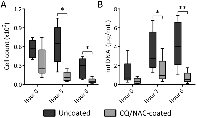
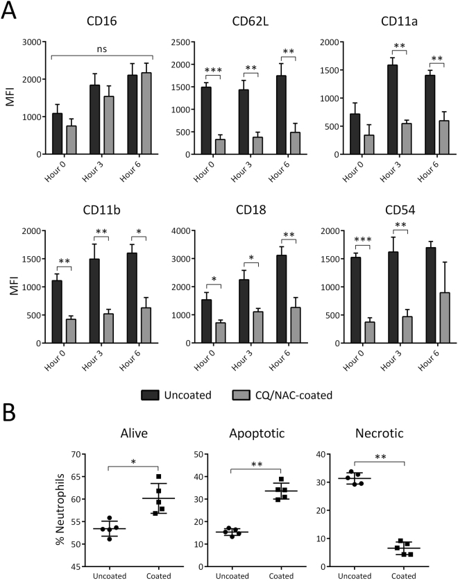
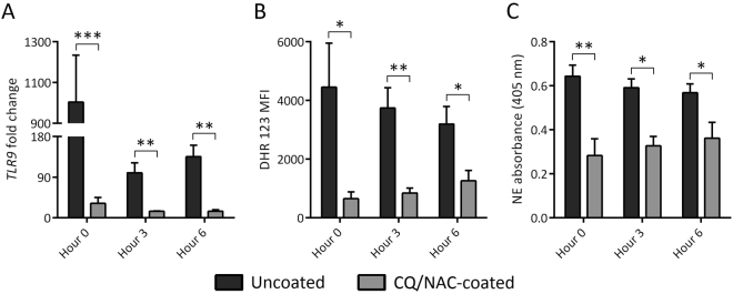
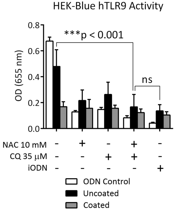
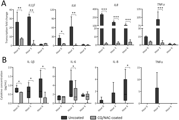
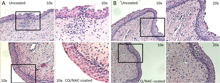
Bladder instrumentation engages the innate immune system via neutrophil activation, promoting inflammation and pain. Elevated levels of mitochondrial DNA (mtDNA) have been associated with tissue damage and organ dysfunction. We hypothesized that local bladder trauma induced by a Foley catheter (FC) will result in mtDNA release, migration of neutrophils into the bladder lumen, and activation of the Toll-like receptor 9 (TLR9) and nuclear factor kappa B (NF-κB) pathway leading to bladder tissue damage. We randomized 10 swine into two groups receiving uncoated, or chloroquine/N-Acetylcysteine (CQ/NAC)-coated FCs. Urine samples were analyzed for mtDNA activation of TLR9/NF-κB as demonstrated by indicators of neutrophil adhesion, migration, and activation. We found that uncoated FCs resulted in a unique active neutrophil phenotype that correlated with bladder epithelial injury, neutrophilia, necrosis, mtDNA release, TLR9/NF-κB activation, transcription and secretion of pro-inflammatory cytokines, and enhanced respiratory burst. In our study we observed that the high levels of mtDNA and elevated TLR9/NF-κB activity were ameliorated in the CQ/NAC-coated FC group. These findings suggest that post-migrated bladder luminal neutrophils are involved in local tissue damage and amelioration of the mtDNA/TLR9/NF-κB inflammatory axis may represent a therapeutic target to prevent inflammation, and bladder tissue injury.


Conflict of interest statement

The authors declare no competing interests.

Figures

Figure 1
Figure 1
High urine neutrophil numbers and mitochondrial DNA concentrations, but low bacterial DNA, found in uncoated Foley catheter (FC) swine. (A) Total live neutrophils were counted by trypan blue exclusion after negative immunomagnetic isolation in both coated (light gray, n = 5) and uncoated (dark gray, n = 5) FC groups. (B) Mitochondrial DNA concentrations were determined by real time PCR using swine Mt-cyb primer and a standard provided by the manufacturer, *p < 0.05, **p < 0.01.
Figure 2
Figure 2
Unique urine neutrophil phenotype correlates with cellular necrosis during Foley catheter (FC)-mediated injury. (A) Fluorescence activated cell sorting (FACS) analysis of active urine CD16/CD62L neutrophils indicating different migration marker phenotypes of CD11a, CD11b, CD18, and CD54. (B) Alive, apoptotic, and necrotic neutrophil percentages were determined by FACS analysis by Annexin V side scatter and 7-Aminoactinomycin D forward scatter gating for both uncoated and coated FC groups. Data shown represents mean MFI + SD, *p < 0.05, **p < 0.01, ***p < 0.001.
Figure 3
Figure 3
Foley catheters (FCs) promote reactive oxygen species (ROS) and neutrophil elastase (NE) activity in a TLR9 dependent manner. (A) Fold change in TLR9 transcription in urine neutrophils from uncoated (dark gray, n = 5) and coated (light gray, n = 5) FC groups as determined by real time PCR against peripheral blood neutrophils. (B) ROS production by these neutrophils in both FC groups at each time point by dihydrorhodamine (DHR) 123 staining fluorescence activated cell sorting (FACS) analysis. (C) Urine neutrophils from both groups were assessed for NE activity after 1-hour incubation with NE-specific substrate by spectrophotometry. Data shown as mean + SD, *p < 0.05, **p < 0.01, ***p < 0.001.
Figure 4
Figure 4
Urine mitochondrial DNA induces TLR9 activity. Urine specimens obtained from uncoated or CQ/NAC coated FC were co-incubated with HEK-Blue™ hTLR9 reporter cell line or pre-incubated with graded concentrations of NAC or CQ alone or TLR9 iODN. After 6 hours supernatants were analyzed for activity by spectroscopy of the target transgene NF-κB-induced secreted embryonic alkaline phosphatase absorbance at 655 nm. Data is representative of triplicate experiments where mean activity + SD was calculated from 5 coated and 5 uncoated FC subjects. HEK – human embryonic kidney; iODN – inhibitory oligodeoxynucleotide; NAC – N-Acetylcysteine; CQ – chloroquine. ***p < 0.001.
Figure 5
Figure 5
Urinary cytokine concentrations and neutrophil cytokine transcription is mediated by chloroquine/N-Acetylcysteine (CQ/NAC) coating. (A) Fold change in transcription of these pro-inflammatory cytokines in urine neutrophils from both FC groups determined real-time qPCR measured against peripheral blood neutrophils, with data shown as mean fold change + SD. (B) Concentrations of pro-inflammatory cytokines found in approximately 20 mL urine from both uncoated (dark gray, n = 5) and coated (light gray, n = 5) Foley catheter (FC) groups determined by enzyme linked immunosorbent assay (ELISA), with data shown as box-and-whisker plots with standard descriptive statistics. *p < 0.05, **p < 0.01, ***p < 0.001.
Figure 6
Figure 6
Coating Foley catheters (FCs) with chloroquine and N-Acetylcysteine promotes transcription of the anti-inflammatory cytokine IL-10. Messenger RNA from urine neutrophils of coated and uncoated FC groups was isolated and assessed for transcription of the anti-inflammatory cytokine IL-10 by real time PCR measured against peripheral blood neutrophil mRNA. Data shown represents results of at least 10 experiments, 5 per group, with mean fold change + SD. **p < 0.01, ***p < 0.001.
Figure 7
Figure 7
Urethra and bladder neck histology from uncoated and coated Foley catheters (FCs). After sample preparation, paraffin-embedded blocks were cut and stained with H&E. Tissue samples at point of contact with uncoated (top) and coated (bottom) FCs in the urethra (A), and bladder neck (B) at 100× magnification with boxes and 200× magnification highlighting distinct morphological and immune cell infiltration differences.
Figure 8
Figure 8
Urethra immunohistochemistry of TLR9 staining from uncoated and coated Foley catheters (FCs). After preparation, paraffin-embedded tissues were deparaffinized and prepared for IHC staining for porcine TLR9 with horseradish peroxidase secondary staining. Tissue samples at point of contact in the urethra for uncoated (A) and CQ/NAC-coated (B) FCs at 200× magnification. Arrows point to notable areas on the epithelial surface and subepithelial layer to show differences in TLR9 expression.

References

    1. Hooton TM, et al. Diagnosis, prevention, and treatment of catheter-associated urinary tract infection in adults. International Clinical Practice Guidelines from the Infectious Diseases Society of America. Clin. Infect. Dis. 2010;50:625–663. doi: 10.1086/650482. - DOI - PubMed
    1. Peychl L, Zalud R. Changes in the urinary bladder caused by short-term permanent catheter insertion. Cas. Lek. Cesk. 2008;147:325–339. - PubMed
    1. Shahin RD, et al. Neutrophil recruitment and bacterial clearance correlated with LPS responsiveness in local gram-negative infection. J. Immunol. 1987;138:3475–3480. - PubMed
    1. Jacobsen SM, et al. Complicated catheter-associated urinary tract infections due to Escherichia coli and Proteus mirabilis. Clin. Microbiol. Rev. 2008;21:26–59. doi: 10.1128/CMR.00019-07. - DOI - PMC - PubMed
    1. Puyo CA, Dahms TE. Innate Immunity Mediating Inflammation Secondary to Endotracheal Intubation. Arch. Otolaryngol. Head Neck Surg. 2012;138:854–858. doi: 10.1001/archoto.2012.1746. - DOI - PubMed

Publication types

MeSH terms