. 2018 Apr 23;8(1):6382.
doi: 10.1038/s41598-018-24904-z.

Tau Internalization is Regulated by 6-O Sulfation on Heparan Sulfate Proteoglycans (HSPGs)

Affiliations

Tau Internalization is Regulated by 6-O Sulfation on Heparan Sulfate Proteoglycans (HSPGs)

Jennifer N Rauch et al. Sci Rep. .

Abstract

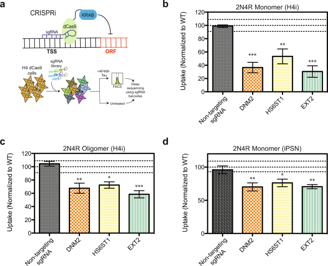
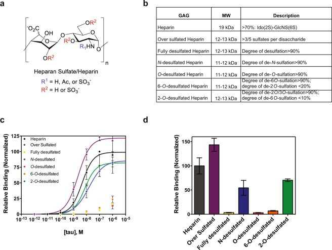
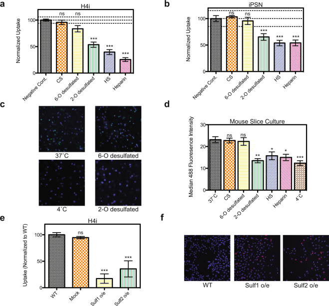
The misfolding and accumulation of tau protein into intracellular aggregates known as neurofibrillary tangles is a pathological hallmark of neurodegenerative diseases such as Alzheimer's disease. However, while tau propagation is a known marker for disease progression, exactly how tau propagates from one cell to another and what mechanisms govern this spread are still unclear. Here, we report that cellular internalization of tau is regulated by quaternary structure and have developed a cellular assay to screen for genetic modulators of tau uptake. Using CRISPRi technology we have tested 3200 genes for their ability to regulate tau entry and identified enzymes in the heparan sulfate proteoglycan biosynthetic pathway as key regulators. We show that 6-O-sulfation is critical for tau-heparan sulfate interactions and that this modification regulates uptake in human central nervous system cell lines, iPS-derived neurons, and mouse brain slice culture. Together, these results suggest novel strategies to halt tau transmission.


Conflict of interest statement

K.S.K. serves as a consultant and has shares in ADRx, serves as co-director of the Tau Consortium and is on the scientific advisory board of Cohen Veterans Bioscience. The remaining authors declare that they have no competing interests.

Figures

Figure 1
Figure 1
Tau uptake is regulated by quaternary structure. (a) 2N4R monomer, oligomer, and fibrillized proteins show distinct banding patterns on a non-reducing SDS-PAGE gel. 2N4R Fibrils before (top) and after (bottom) sonication as visualized by negative-stain EM. Bar represents 500 nm (b) DLS of 2N4R monomer, oligomer, fibril, and sonicated fibril species. Experiments were performed in triplicate and the error shown is SD. (c) Uptake of 2N4R quaternary structures in H4 cells after 1 h at 37 °C. (d) Uptake of 2N4R quaternary structures in iPS-derived neurons after 1 h at 37 °C. For all uptake experiments, three independent experiments were performed in duplicate, identical control experiments were performed at 4 °C and subtracted from 37 °C data to generate final curves shown, and the error shown is SEM.
Figure 2
Figure 2
CRISPRi screen for tau uptake modulators. (a) Screening strategy using H4 CRISPRi cells, see text for details. (b) Reconfirmation of selected hits in H4i cells with 2N4R monomer (50 nM, 1 h at 37 °C), normalized to a WT (no sgRNA) control. (c) Uptake of 2N4R oligomers (50 nM, 1 h at 37 °C) in H4i cells with selected gene knockdowns, normalized to a WT (no sgRNA) control. (d) Uptake of 2N4R monomer (200 nM, 1 h at 37 °C) in iPS-derived neurons with selected gene knockdowns, normalized to a WT (no sgRNA) control. All uptake experiments were performed in duplicate over three independent experiments with the data combined. Lines on the graphs represent WT mean +/− 3 standard deviations, error bars represent SEM, a one-way ANOVA analysis with Dunnett’s method was used to determine significance between the gene knockdown and the non-targeting sgRNA *p-value ≤ 0.05, **p-value ≤ 0.01, ***p-value ≤ 0.001.
Figure 3
Figure 3
Binding of tau to heparin derivatives. (a) HS chains consist of GlcA/IdoA-GlcNAc disaccharide units that can be modified at positions indicated in red and blue. (b) List of heparin/heparin derivatives that were used, their average molecular weights, and description of their sulfation modifications. (c) Binding of 2N4R to various heparin derivatives by ELISA with data fit to a Hill binding model (line) where appropriate. (d) Normalized relative binding of 2N4R (10 nM) to various heparin derivatives. Three independent experiments were performed, data were normalized to heparin controls, and the error shown is SD.
Figure 4
Figure 4
6-O sulfation regulates tau uptake in CNS culture. (a) Internalization of 2N4R-488 (50 nM, 1 h at 37 °C) into H4 cells is strongly inhibited by incubation with heparin, heparan sulfate, and 2-O-desulfated heparin as compared to 6-O-desulfated heparin or chondroitin sulfate (0.05 mg/ml for all derivatives). (b) Internalization of 2N4R-488 (200 nM, 1 h at 37 °C) into iPS-derived neurons is inhibited by incubation with heparin, heparan sulfate, and 2-O-desulfated heparin as compared to 6-O-desulfated heparin or chondroitin sulfate (0.5 mg/ml for all derivatives). All uptake experiments were performed in duplicate over three independent experiments with the data combined. Lines on the graphs represent WT mean +/− 3 standard deviations, error bars represent SEM, and a one-way ANOVA analysis with Dunnett’s method was used to determine significance compared to the negative control *p-value ≤ 0.05, **p-value ≤ 0.01, ***p-value ≤ 0.001, ns = not significant. (c) Internalization of 2N4R-488 (200 nM, 30 min at 37 °C) into mouse slice culture is strongly inhibited by incubation with 2-O-desulfated heparin, but not 6-O-desulfated heparin (0.5 mg/ml). Hoechst stain is used to label nuclei. (d) Quantification of the median 488 fluorescence intensity for tau uptake in mouse slice culture. Two independent experiments were performed with multiple images (>5) from each condition. Error bars represent SEM. (e) Uptake of 2N4R-488 (50 nM, 1 h at 37 °C) tau is reduced when Sulf1 or Sulf2 is overexpressed in H4 cells as compared to WT or mock transfected cells. Uptake experiments were performed in duplicate over three independent experiments with the data normalized to WT and combined. Error bars represent SEM. (f) ICC confirms that Sulf1 and Sulf2 are overexpressed (red) in H4 cells. Hoechst stain is used to label nuclei.

References

    1. Brandt R, Hundelt M, Shahani N. Tau alteration and neuronal degeneration in tauopathies: mechanisms and models. Biochim Biophys Acta. 2005;1739:331–354. doi: 10.1016/j.bbadis.2004.06.018. - DOI - PubMed
    1. Jeganathan S, von Bergen M, Mandelkow EM, Mandelkow E. The natively unfolded character of tau and its aggregation to Alzheimer-like paired helical filaments. Biochemistry. 2008;47:10526–10539. doi: 10.1021/bi800783d. - DOI - PubMed
    1. Schweers O, Schonbrunn-Hanebeck E, Marx A, Mandelkow E. Structural studies of tau protein and Alzheimer paired helical filaments show no evidence for beta-structure. J Biol Chem. 1994;269:24290–24297. - PubMed
    1. Hyman BT, Van Hoesen GW, Damasio AR, Barnes CL. Alzheimer’s disease: cell-specific pathology isolates the hippocampal formation. Science. 1984;225:1168–1170. doi: 10.1126/science.6474172. - DOI - PubMed
    1. Braak H, Braak E. Neuropathological stageing of Alzheimer-related changes. Acta neuropathologica. 1991;82:239–259. doi: 10.1007/BF00308809. - DOI - PubMed

Publication types

MeSH terms