. 2018 Apr 9:9:486.
doi: 10.3389/fimmu.2018.00486. eCollection 2018.

CD161 Defines a Functionally Distinct Subset of Pro-Inflammatory Natural Killer Cells

Collaborators, Affiliations

CD161 Defines a Functionally Distinct Subset of Pro-Inflammatory Natural Killer Cells

Ayako Kurioka et al. Front Immunol. .

Abstract

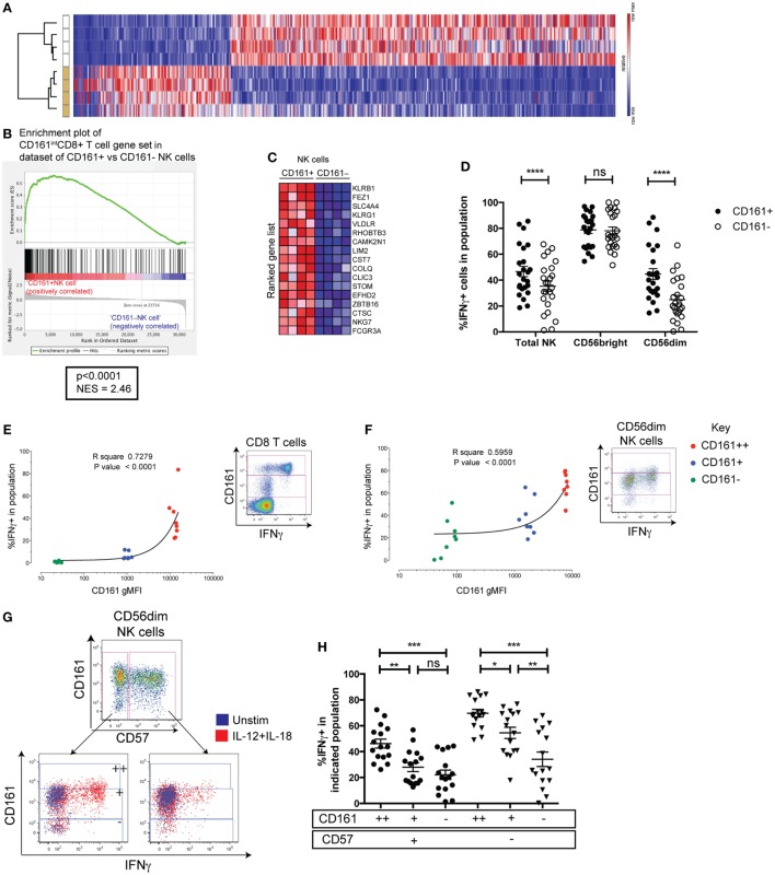
CD161 is a C-type lectin-like receptor expressed on the majority of natural killer (NK) cells; however, the significance of CD161 expression on NK cells has not been comprehensively investigated. Recently, we found that CD161 expression identifies a transcriptional and innate functional phenotype that is shared across various T cell populations. Using mass cytometry and microarray experiments, we demonstrate that this functional phenotype extends to NK cells. CD161 marks NK cells that have retained the ability to respond to innate cytokines during their differentiation, and is lost upon cytomegalovirus-induced maturation in both healthy and human immunodeficiency virus (HIV)-infected patients. These pro-inflammatory NK cells are present in the inflamed lamina propria where they are enriched for integrin CD103 expression. Thus, CD161 expression identifies NK cells that may contribute to inflammatory disease pathogenesis and correlates with an innate responsiveness to cytokines in both T and NK cells.

Keywords: CD161; cytomegalovirus; human immunodeficiency virus; inflammatory bowel diseases; natural killer cells; pro-inflammatory cytokines.


Figures

Figure 1
Figure 1
CD161 expression defines two distinct subsets of natural killer (NK) cells. (A) Gating strategy used to identify CD161+ and CD161 NK cells. (B) Representative staining showing the expression of CD161 in umbilical cord blood, 24-month olds (24mo), and adult individuals on total NK cells (CD3CD56+ cells). (C) Frequencies of CD161+NK cells in cord blood (n = 5), 24-month olds (n = 28), and adult individuals (n = 33). (D) Representative staining showing the expression of CD161 on CD56bright and CD56dimNK cells in adult peripheral blood. (E) Frequencies of CD161+ cells within CD56bright and CD56dimNK cells (n = 33).
Figure 2
Figure 2
CD161+NK cells share high responsiveness to IL-12+IL-18 with CD161-expressing T cells. (A) Heatmap illustrating significantly differentially expressed transcripts between CD161+ and CD161 NK cells in four donors. Subsets clustered by one minus Pearson correlation by GENE-E. (B) Gene set enrichment analysis of CD161+ vs. CD161 NK cells, showing a significant enrichment of genes upregulated in CD161+(intermediate) (CD161int) CD8+T cells compared to CD161CD8+T cells (red = positively correlated, blue = negatively correlated) within CD161+ NK cells compared to CD161 NK cells. (C) Heatmap shows top 17 genes that are highly (red) or lowly (blue) expressed in either CD161+ or CD161 NK cells. NES = normalized enrichment score. (D) IFNγ production by CD161+ and CD161 NK cells in response to overnight IL-12+IL-18 stimulation. N = 24. (E,F) gMFI of CD161 for CD161bright (++) (red), CD161int(+) (blue), and CD161 (green) subsets in CD8+T cells (E) or CD56dimNK cells (F), correlated with IFNγ expression in response to IL-12+IL-18 (n = 9). Representative staining showing IFNγ expression on indicated cells from the same donor, and gating of CD161bright (++), CD161int(+), and CD161 subsets based on CD8+T cells, are shown. (G) Representative staining showing IFNγ production in response to IL-12+IL-18 from CD57+ and CD57 cells within the CD56dim natural killer (NK) cell population (left). CD161bright (++), CD161int(+), and CD161 subsets were gated within CD57+ and CD57 NK cells as indicated. (H) Frequency of IFNγ+ cells in each population was graphed.
Figure 3
Figure 3
CD161+ natural killer (NK) cells have a greater proliferative capacity than CD161 NK cells. Peripheral blood mononuclear cells from healthy donors were CTV-labeled and cultured for 5 days with the indicated stimuli. (A) Representative staining of CellTrace Violet (CTV) for CD161+ and CD161 NK cells from one healthy donor. (B,C) Frequency of CD161+ and CD161 NK cells either (B) diluting CTV, or (C) expressing CD69, after simulation with the indicated combinations of cytokines and PHA (n = 14). Graphs show frequencies of CTVlow and CD69+ cells following background subtraction. (D) Frequency of CD161+ and CD161 NK cells expressing phosphatidylserine after 5 days culture with the indicated stimuli (n = 4). (E,F) CTV-labeled FACS-sorted CD161+ and CD161 NK cells were stimulated with IL-15 for 5 days or left unstimulated, and the frequency of cells diluting CTV (CTVlow) was analyzed (n = 3). (E) Representative staining and (F) frequency data are shown. (G) Representative staining for CTV on CD57+ and CD57 NK cells, according to CD161 expression. (H) Frequency of cells diluting CTV on indicated CD56dim NK cell subsets stimulated with IL-15, according to CD161 and CD57 expression (n = 3). CD56brightNK cells were excluded as they do not express CD57.
Figure 3
Figure 3
CD161+ natural killer (NK) cells have a greater proliferative capacity than CD161 NK cells. Peripheral blood mononuclear cells from healthy donors were CTV-labeled and cultured for 5 days with the indicated stimuli. (A) Representative staining of CellTrace Violet (CTV) for CD161+ and CD161 NK cells from one healthy donor. (B,C) Frequency of CD161+ and CD161 NK cells either (B) diluting CTV, or (C) expressing CD69, after simulation with the indicated combinations of cytokines and PHA (n = 14). Graphs show frequencies of CTVlow and CD69+ cells following background subtraction. (D) Frequency of CD161+ and CD161 NK cells expressing phosphatidylserine after 5 days culture with the indicated stimuli (n = 4). (E,F) CTV-labeled FACS-sorted CD161+ and CD161 NK cells were stimulated with IL-15 for 5 days or left unstimulated, and the frequency of cells diluting CTV (CTVlow) was analyzed (n = 3). (E) Representative staining and (F) frequency data are shown. (G) Representative staining for CTV on CD57+ and CD57 NK cells, according to CD161 expression. (H) Frequency of cells diluting CTV on indicated CD56dim NK cell subsets stimulated with IL-15, according to CD161 and CD57 expression (n = 3). CD56brightNK cells were excluded as they do not express CD57.
Figure 4
Figure 4
CD161+ NK cells are enriched for CD103-expressing cells in the inflamed gut. (A,B) FACS plots (A) and bar graph (B) showing CD103 expression on indicated natural killer (NK) cell populations isolated from the periphery (n = 5), tonsil (n = 1), or the lamina propria, either inflamed from IBD patients (n = 7) or non-inflamed lamina propria (n = 4) as analyzed by mass cytometry. FACS plots gated on total CD3CD56+ NK cells. IBD, inflammatory bowel disease.
Figure 5
Figure 5
Changes in CD161 expression in cytomegalovirus (CMV) and human immunodeficiency virus (HIV) infection. (A) Frequencies of CD161 NK cells within the CD56dim natural killer (NK) cell population within CMV+ (n = 16) and CMV− (n = 16) individuals. Data combined from four independent experiments. (B,C) Frequencies of (B) CD57+ cells or (C) NKG2C+ cells in CD161+ or CD161 population of CD56dim NK cells, in CMV+ and CMV− donors. N = 14 for CMV+, n = 11 for CMV− individuals. (D) Peripheral and cord blood samples were stimulated with IL-12+IL-18 overnight and acquired by mass cytometry. (H) t-distributed stochastic neighbor embedding analysis was performed on CMV+ (n = 5) and CMV− donors (n = 6). The median intensities of each marker were calculated for the NK cell populations identified (left) and plotted as a heatmap (right). (E–L) The frequency of CD161-expressing NK cells in patients with chronic-stage HIV infection from the Swiss HIV cohort study, who were followed for 2 years of anti-retroviral therapy (ART), with samples taken prior to the start of the treatment (T-0), at 1 year (T-1), and 2 years (T-2) into treatment. Patients were tested for CMV seropositivity prior to the start of treatment (n = 27, Table S2 in Supplementary Material). The frequency of CD161+ NK cells within (E) total NK cells (CD3CD56+), (F) CD56dim NK cells, and (G) CD56bright NK cells is shown for HIV patients at T-0 (n = 25 for CMV+, n = 2 for CMV−), compared to CMV+ (n = 16) and CMV− (n = 16) healthy donors. (H–J) Frequency of CD161+ NK cells within HIV patients followed for 2 years of ART. CD161+ cells within (H) total NK cells, (I) CD56dim NK cells, and (J) CD56bright NK cells are shown for HIV patients at T-0, T-1, and T-2. (K) Representative staining showing CD161 expression on total NK cells from one patient at T-0 (left), T-1 (center), and T-2 (right). (L) Histogram overlaying CD161 expression on total NK cells from one patient.

References

    1. Spits H, Bernink JH, Lanier L. NK cells and type 1 innate lymphoid cells: partners in host defense. Nat Immunol (2016) 17:758–64.10.1038/ni.3482 - DOI - PubMed
    1. Bennett IM, Zatsepina O, Zamai L, Azzoni L, Mikheeva T, Perussia B. Definition of a natural killer NKR-P1A+/CD56-/CD16- functionally immature human NK cell subset that differentiates in vitro in the presence of interleukin 12. J Exp Med (1996) 184:1845–56.10.1084/jem.184.5.1845 - DOI - PMC - PubMed
    1. Konjević G, Mirjacić Martinović K, Vuletić A, Jurisić V, Spuzić I. Distribution of several activating and inhibitory receptors on CD3-CD16+ NK cells and their correlation with NK cell function in healthy individuals. J Membr Biol (2009) 230:113–23.10.1007/s00232-009-9191-3 - DOI - PubMed
    1. Aldemir H, Prod’homme V, Dumaurier M-J, Retiere C, Poupon G, Cazareth J, et al. Cutting edge: lectin-like transcript 1 is a ligand for the CD161 receptor. J Immunol (2005) 175:7791–5.10.4049/jimmunol.175.12.7791 - DOI - PubMed
    1. Rosen DB, Bettadapura J, Alsharifi M, Mathew PA, Warren HS, Lanier LL. Cutting edge: lectin-like transcript-1 is a ligand for the inhibitory human NKR-P1A receptor. J Immunol (2005) 175:7796–9.10.4049/jimmunol.175.12.7796 - DOI - PubMed

Publication types