Case Reports
. 2018 Mar 4:2018:9278526.
doi: 10.1155/2018/9278526. eCollection 2018.

A Seizure and Hemiplegia following Contrast Exposure: Understanding Contrast-Induced Encephalopathy

Affiliations
Case Reports

A Seizure and Hemiplegia following Contrast Exposure: Understanding Contrast-Induced Encephalopathy

Bhargavi Donepudi et al. Case Rep Med. .

Abstract

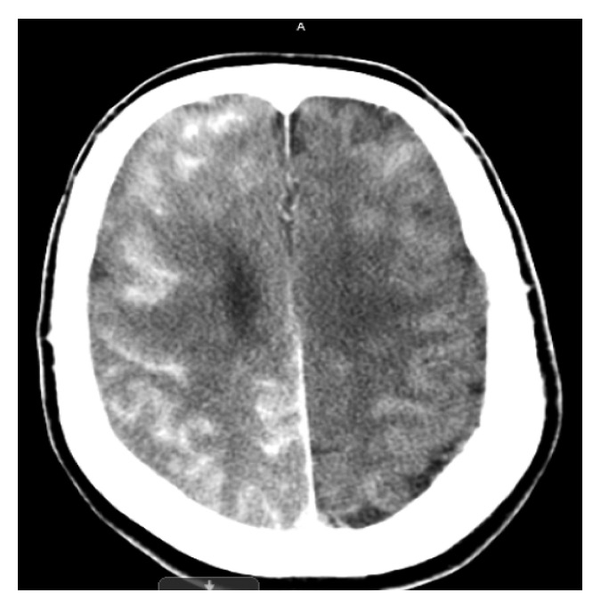
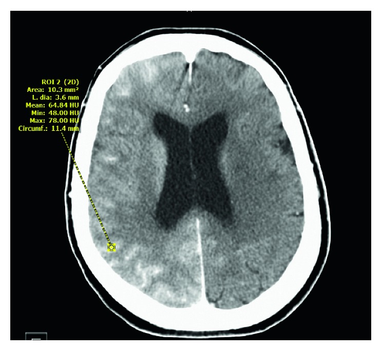
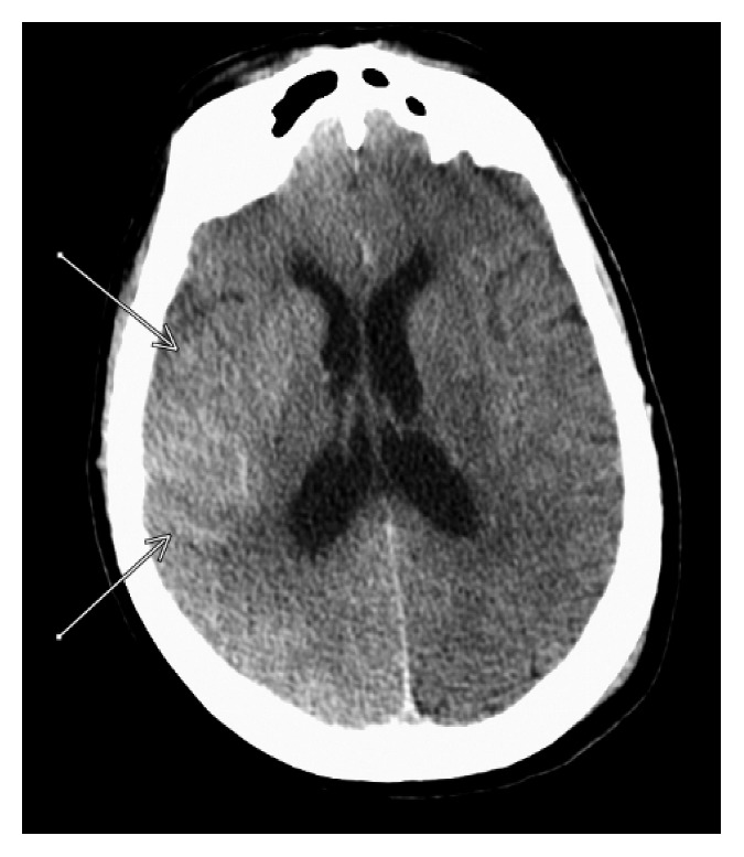
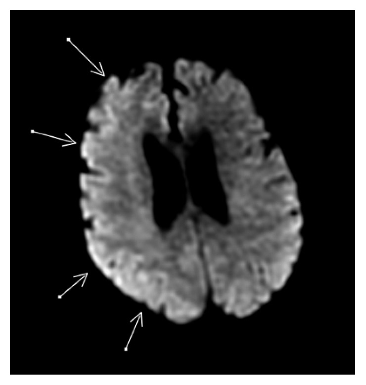
Contrast-induced encephalopathy is a rare, reversible phenomenon known to occur after intravenous or intra-arterial contrast exposure. This report describes a case involving a 73-year-old female admitted for an elective thoracic aortic aneurysm repair. During the procedure, a large volume of nonionic iodinated contrast was necessary for arteriography. Postoperatively, the patient developed seizure activity followed by left-sided hemiplegia. Computed tomography (CT) of the brain without contrast and magnetic resonance imaging (MRI) were negative for acute stroke but did show residual contrast surrounding the brain. Antiepileptic medications were administered with resolution of the seizure activity. The patient was treated with supportive management and improved to baseline over the next seven days. This case demonstrates a rare, nonionic iodinated contrast-induced encephalopathy with seizure activity and transient hemiplegia. The unique imaging findings differentiate it from other neurologic conditions.


Figures

Figure 1
Figure 1
CT brain showing increased hyperdensity and enhancement of right cortex.
Figure 2
Figure 2
Measurement of Hounsfield units (HU) which differentiate blood (30–45 HU) from contrast material (80–160). It shows a mean of 64 HU.
Figure 3
Figure 3
CT of the brain on POD one showed decreased hyperintensity on the right side of the cortex compared to the CT of the brain immediately post-op (Figure 1).
Figure 4
Figure 4
MRI with DWI showing diffuse involvement of the frontoparietal region.

Similar articles

Cited by

References

    1. Yan J., Ramanathan V. Severe encephalopathy following cerebral arteriogram in a patient with end-stage renal disease. Seminars in Dialysis. 2013;26(2):203–207. doi: 10.1111/sdi.12061. - DOI - PubMed
    1. Yu J., Dangas G. New insights into the risk factors of contrast-induced encephalopathy. Journal of Endovascular Therapy. 2011;18(4):545–546. doi: 10.1583/11-3476c.1. - DOI - PubMed
    1. Iwata T., Mori T., Tajiri H., Miyazaki Y., Nakazaki M. Repeated injections of contrast medium inducing dysfunction of the blood-brain barrier. Neurologia Medico-Chirurgica. 2013;53(1):34–36. doi: 10.2176/nmc.53.34. - DOI - PubMed
    1. Law S., Panichpisal K., Demede M., et al. Contrast-induced neurotoxicity following cardiac catheterization. Case Reports in Medicine. 2012;2012:4. doi: 10.1155/2012/267860.267860 - DOI - PMC - PubMed
    1. Spina R., Simon N., Markus R., Muller D. W. M., Kathir K. Contrast-induced encephalopathy following cardiac catheterization. Catheterization and Cardiovascular Interventions. 2016;90(2):257–268. doi: 10.1002/ccd.26871. - DOI - PubMed

Publication types

LinkOut - more resources