. 2018 Mar 4:2018:6079642.
doi: 10.1155/2018/6079642. eCollection 2018.

Human Umbilical Cord MSC-Derived Exosomes Suppress the Development of CCl4-Induced Liver Injury through Antioxidant Effect

Affiliations

Human Umbilical Cord MSC-Derived Exosomes Suppress the Development of CCl4-Induced Liver Injury through Antioxidant Effect

Wenqian Jiang et al. Stem Cells Int. .

Abstract

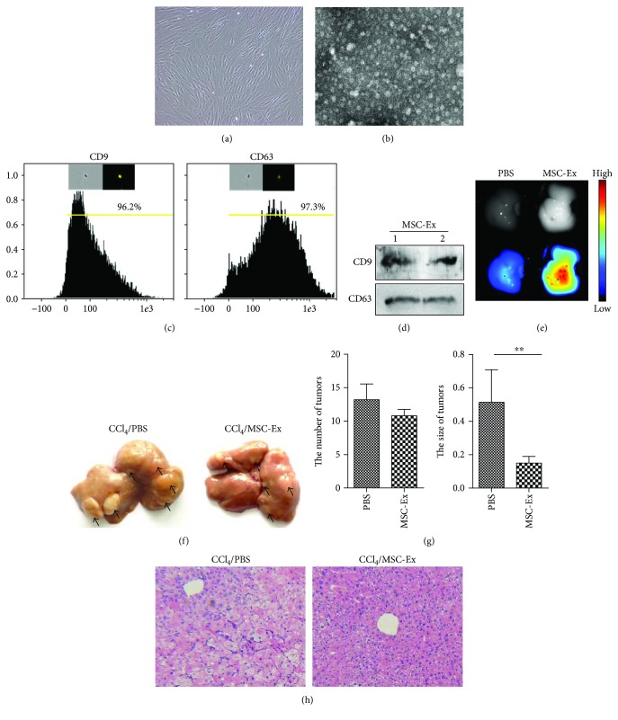
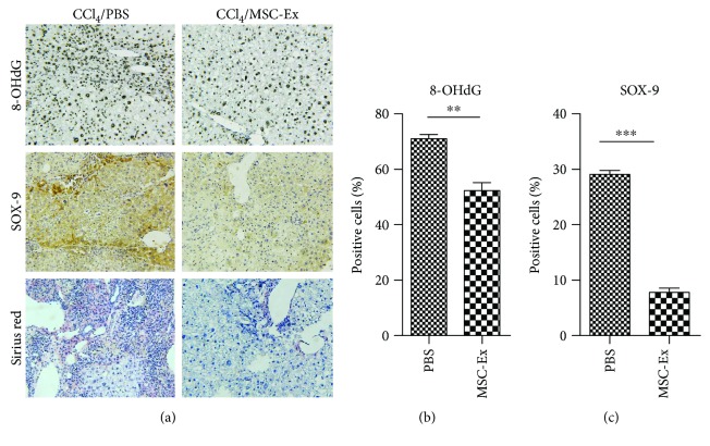
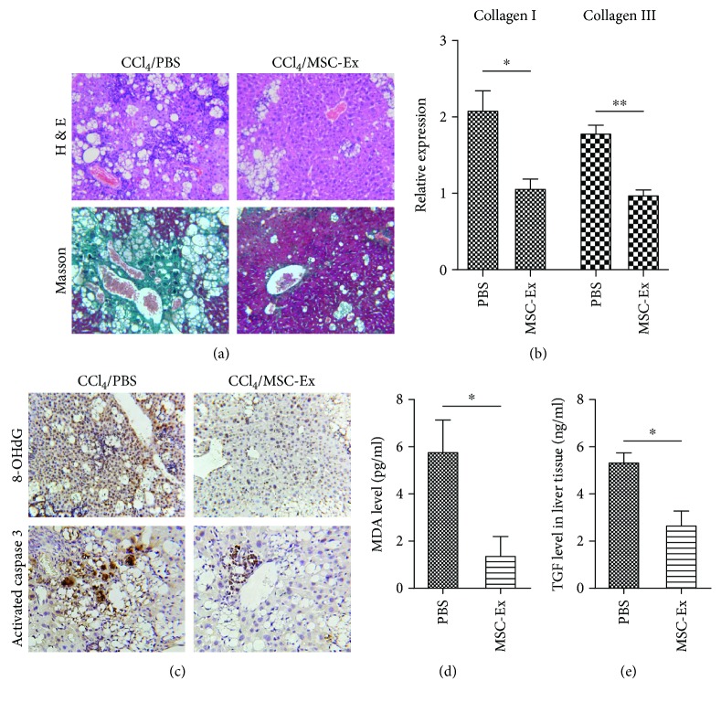
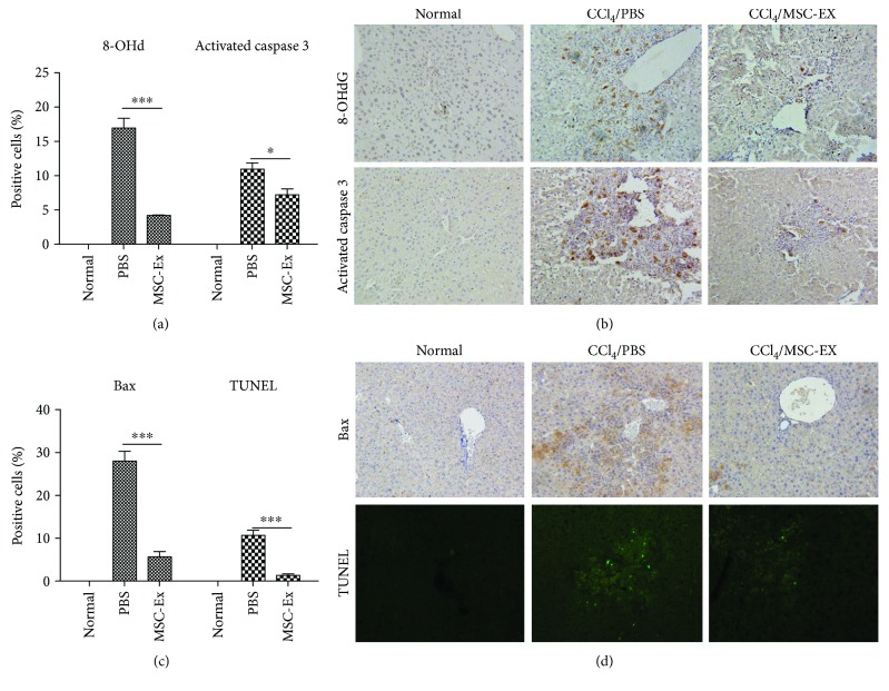
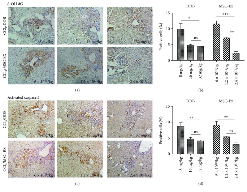
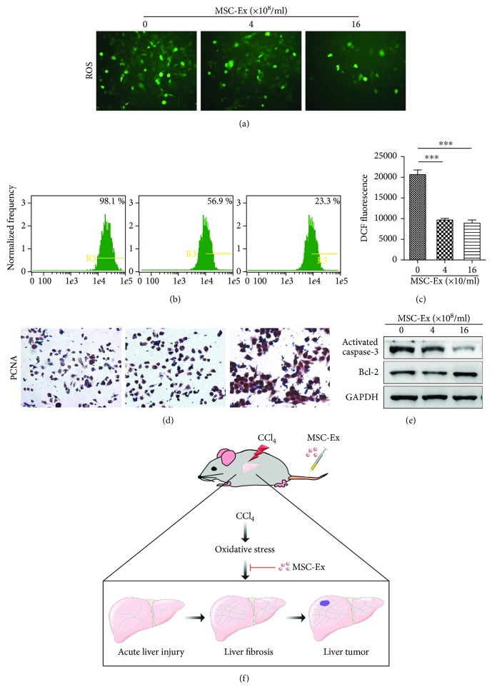
Mesenchymal stem cells (MSCs) have been increasingly applied into clinical therapy. Exosomes are small (30-100 nm in diameter) membrane vesicles released by different cell types and possess the similar functions with their derived cells. Human umbilical cord MSC-derived exosomes (hucMSC-Ex) play important roles in liver repair. However, the effects and mechanisms of hucMSC-Ex on liver injury development remain elusive. Mouse models of acute and chronic liver injury and liver tumor were induced by carbon tetrachloride (CCl4) injection, followed by administration of hucMSC-Ex via the tail vein. Alleviation of liver injury by hucMSC-Ex was determined. We further explored the production of oxidative stress and apoptosis in the development of liver injury and compared the antioxidant effects of hucMSC-Ex with frequently used hepatic protectant, bifendate (DDB) in liver injury. hucMSC-Ex alleviated CCl4-induced acute liver injury and liver fibrosis and restrained the growth of liver tumors. Decreased oxidative stress and apoptosis were found in hucMSC-Ex-treated mouse models and liver cells. Compared to bifendate (DDB) treatment, hucMSC-Ex presented more distinct antioxidant and hepatoprotective effects. hucMSC-Ex may suppress CCl4-induced liver injury development via antioxidant potentials and could be a more effective antioxidant than DDB in CCl4-induced liver tumor development.


Figures

Figure 1
Figure 1
hucMSC-Ex suppressed CCl4-induced mouse liver tumor growth. (a) Morphological appearance of cultured hucMSCs. Original magnification 200x. (b) Identification of hucMSC-Ex with transmission electron micrograph. Scale bar 100 nm. (c) Imaging flow cytometer analysis of phenotypic markers of hucMSC-Ex. hucMSC-Ex were positive for CD9 and CD63. (d) Western blot assay indicated the positive expression of CD9 and CD63 proteins in hucMSC-Ex. (e) Distribution of CM-DiR labeled hucMSC-Ex in CCl4-induced hepatic carcinoma mice by in vivo fluorescent imaging. (f) Macroscopical observation of tumors in the liver surface of mice treated with PBS or hucMSC-Ex. The arrows indicated the tumors. (g) Analysis of the number and average size of tumors in the livers. Tumor size was significantly reduced in the hucMSC-Ex group (n = 6; ∗∗P < 0.01). (h) Representative images of H&E staining in the livers treated with PBS or hucMSC-Ex. Original magnification 100x.
Figure 2
Figure 2
hucMSC-Ex reduced oxidative stress in mouse liver tumor. (a, b) The levels of 8-OHdG, SOX9, and collagen in mouse livers were measured by immunohistochemistry or Sirius red staining in liver tumor treated with PBS or hucMSC-Ex. Original magnification 200x. Compared with the PBS group, hucMSC-Ex significantly reduced production of oxidative stress marker 8-OHdG, SOX9 expression, and collagen deposition in liver tumor (n = 6; ∗∗P < 0.01, ∗∗∗P < 0.001).
Figure 3
Figure 3
hucMSC-Ex reduced oxidative stress in mouse liver fibrosis. (a) Representative images of H&E and Masson staining of mouse fibrotic livers after PBS or hucMSC-Ex treatment. Reduced steatosis and collagen deposition were observed in the hucMSC-Ex group. Original magnification 200x. (b) Quantitative analyses of collagen I and III mRNA expression after hucMSC-Ex treatment (n = 10; P < 0.05, ∗∗P < 0.01). (c) Immunohistochemistry analysis of 8-OHdG and activated caspase 3 after administration of PBS or hucMSC-Ex. Original magnification 200x. (d, e) MDA and TGF-β levels were measured in homogenates of a mouse fibrotic liver treated with PBS and hucMSC-Ex. The levels of MDA and TGF-β were inhibited by hucMSC-Ex compared with the PBS group (n = 10; P < 0.05).
Figure 4
Figure 4
hucMSC-Ex reduced oxidative stress in acute liver injury. (a, b) Immunohistochemistry staining of 8-OHdG and activated caspase 3 in mouse livers. Reduced production of oxidative stress marker 8-OHdG and activated caspase 3 expression was detected in the hucMSC-Ex group (n = 10; P < 0.05, ∗∗∗P < 0.001). Original magnification 200x. (c, d) Immunohistochemistry staining of Bax and TUNEL in mouse liver slides. Bax expression and cell apoptosis were decreased in the hucMSC-Ex group (n = 10; ∗∗∗P < 0.001). Original magnification 200x.
Figure 5
Figure 5
hucMSC-Ex exerted a more effective antioxidant than DDB in CCl4-induced liver injury. (a, b) Immunohistochemistry staining of 8-OHdG in mouse livers after treatment with DDB (8 mg/kg, 16 mg/kg, and 32 mg/kg) or hucMSC-Ex (6 × 1010 particles/kg, 1.2 × 1011 particles/kg, and 2.4 × 1011 particles/kg) (n = 10; P < 0.05, ∗∗P < 0.01, and ∗∗∗P < 0.001). (c, d) Immunohistochemistry staining of activated caspase 3 in mouse livers after treatment with DDB (8 mg/kg, 16 mg/kg, and 32 mg/kg) or hucMSC-Ex (6 × 1010 particles/kg, 1.2 × 1011 particles/kg, and 2.4 × 1011 particles/kg) (n = 10; ∗∗P < 0.01). Compared with the PBS group, hucMSC-Ex (2.4 × 1011 particles/kg) exerted a more distinct inhibition effect in the expression of 8-OHdG and activated caspase 3. Original magnification 200x.
Figure 6
Figure 6
hucMSC-Ex inhibits oxidative stress in CCl4-injured L02 cells. (a) ROS production in CCl4-injured L02 cells treated with different doses of hucMSC-Ex (0 particles/ml, 4 × 108 particles/ml, and 16 × 108 particles/ml). Original magnification 200x. (b) Percentage of DCF-positive hepatocytes in hucMSC-Ex treated L02 cells using imaging flow cytometer. Data showed that hucMSC-Ex significantly decreased percentage of DCF-positive hepatocytes. (c) Fluorescence intensity of DCF-positive L02 cells was significantly decreased by hucMSC-Ex treatment. DCF fluorescent values are means ± SD. (n = 3; ∗∗∗P < 0.001). (d) Immunohistochemistry staining of PCNA in hucMSC-Ex-treated L02 cells. Percentage of PCNA-positive L02 cells and PCNA expression was enhanced in the hucMSC-Ex group compared with that in the PBS group. Original magnification 200x. (e) Western blot quantification of activated caspase 3 and Bcl2. hucMSC-Ex induced Bcl2 expression and inhibited activated caspase 3 expression in CCl4-injured L02 cells. (f) Experimental model design of hucMSC-Ex in CCl4-induced liver tumor development.

Similar articles

Cited by

References

    1. Cichoz-Lach H., Michalak A. Oxidative stress as a crucial factor in liver diseases. World Journal of Gastroenterology. 2014;20(25):8082–8091. doi: 10.3748/wjg.v20.i25.8082. - DOI - PMC - PubMed
    1. Takaki A., Yamamoto K. Control of oxidative stress in hepatocellular carcinoma: helpful or harmful? World Journal of Hepatology. 2015;7(7):968–979. doi: 10.4254/wjh.v7.i7.968. - DOI - PMC - PubMed
    1. Squillaro T., Peluso G., Galderisi U. Clinical trials with mesenchymal stem cells: an update. Cell Transplantation. 2016;25(5):829–848. doi: 10.3727/096368915X689622. - DOI - PubMed
    1. Anjos-Afonso F., Bonnet D. Nonhematopoietic/endothelial SSEA-1+ cells define the most primitive progenitors in the adult murine bone marrow mesenchymal compartment. Blood. 2007;109(3):1298–1306. doi: 10.1182/blood-2006-06-030551. - DOI - PubMed
    1. In 't Anker P. S., Scherjon S. A., Kleijburg-van der Keur C., et al. Isolation of mesenchymal stem cells of fetal or maternal origin from human placenta. Stem Cells. 2004;22(7):1338–1345. doi: 10.1634/stemcells.2004-0058. - DOI - PubMed

LinkOut - more resources