. 2018 Apr 16:15:28.
doi: 10.1186/s12986-018-0265-z. eCollection 2018.

Energy restriction, exercise and atorvastatin treatment improve endothelial dysfunction and inhibit miRNA-155 in the erectile tissue of the aged rat

Affiliations

Energy restriction, exercise and atorvastatin treatment improve endothelial dysfunction and inhibit miRNA-155 in the erectile tissue of the aged rat

B Rocha et al. Nutr Metab (Lond). .

Abstract

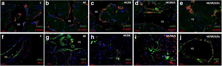
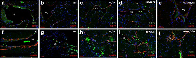
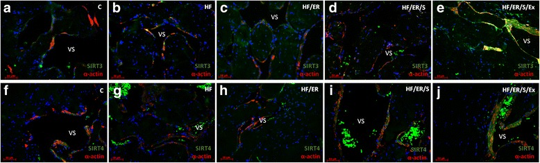
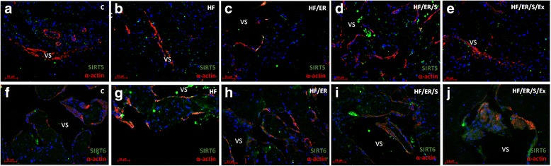
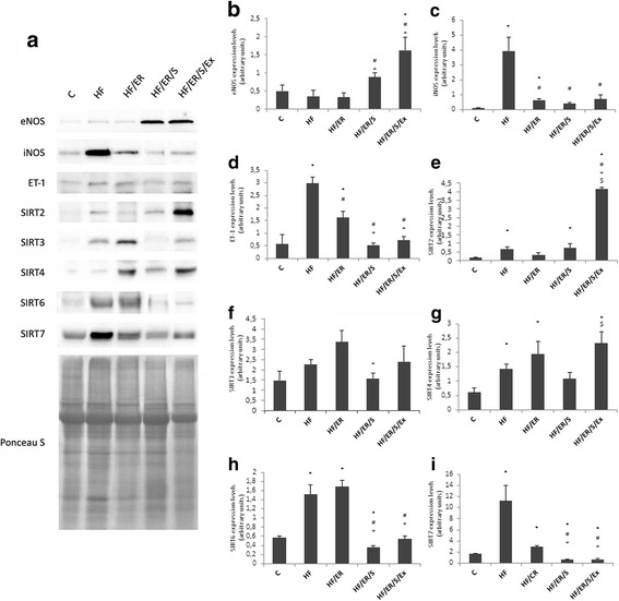
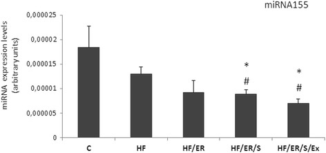
Background: Endothelial dysfunction underlies cardiovascular disease that frequently affects aged individuals. Characterized by local decrease in nitric oxide, it results from down-regulation of endothelial nitric oxide synthase (eNOS) expression/activity. Aiming to elucidate the molecular mechanisms involved in age-related endothelial dysfunction and to unveil potential therapeutic targets, we tested how diet pattern, exercise and atorvastatin modulate the expression of eNOS, inducible NOS (iNOS), endothelin-1, sirtuins (SIRT) and microRNA-155 in the erectile tissue of high-fat fed aged rats.

Methods: Sprague-Dawley male rats fed with high-fat diet until they completed 12 months were grouped and subjected to energy restriction (ER), ER and atorvastatin, or, ER, atorvastatin and physical exercise. Controls were fed with standard rodent chow. The blood pressure was measured using the tail-cuff method before sacrifice at 18 months. Glucose, total cholesterol, HDL, triglyceride and CRP were assessed in blood and eNOS, endothelin-1, iNOS and sirtuins were detected by immunofluorescence in the penis sections; eNOS, endothelin-1, iNOS, SIRT2-4 and SIRT6-7 were semi-quantified by western blotting in tissue homogenates. MicroRNA-155 was quantified using RT-PCR in formalin-fixed paraffin embedded sections. To compare the studied variables, two-tail student t test was used.

Results: Atorvastatin promotes eNOS expression and is more efficient than ER or exercise in the control of hyperlipidemia and inflammation. Among the studied sirtuins, detected for the first time in the erectile tissue of the aged rat, SIRT2 aligns with eNOS expression. Both proteins exhibit over-expression in animals with combined exercise, atorvastatin and ER. Analysis of microRNA-155 expression also suggests its intervention in the regulation of eNOS expression. ER, particularly when combined with atorvastatin, was able to reverse the increase of iNOS and endothelin-1 in high-fat fed rats.

Conclusions: The present results indicate that the association of ER, atorvastatin and exercise is more efficient than isolated interventions in the prevention of endothelial dysfunction.

Keywords: Atorvastatin; Endothelial dysfunction; Energy restriction; Exercise; Sirtuins; microRNA-155.


Conflict of interest statement

All animals procedures were undertaken according to the European Community Guidelines (86/ 609/ EEC) and the Portuguese Act (129/ 92) for the use of experimental animals.All authors approved the final version of this manuscript.All authors declare that they have no competing interest.Springer Nature remains neutral with regard to jurisdictional claims in published maps and institutional affiliations.

Figures

Fig. 1
Fig. 1
Dual immunolabeling of eNOS/α-actin (a-e) and iNOS/ET1 (f-j) in erectile tissue of rats from all experimental groups (n = 3/group). Smooth muscle cells (SMC) layer was evident after α-actin labeling (red), surrounding endothelium (not labeled) and vascular spaces in all animals. No marked differences among groups were evident. eNOS was detected not only in the endothelium, but also in SMC (green) apparently with higher intensity in HF/ER, HF/ER/S and HF/ER/S/Ex groups (c-e). iNOS was detected in the SMC (green) in all experimental groups (f-j), being particularly evident in HF-treated rats (g). ET-1 was mostly localized in the endothelium (red), with an apparent higher intensity in HF-fed rats (g). C-control; HF-high-fat diet treated rats; HF/ER-high-fat diet treated rats under energy restriction for 6 months; HF/ER/S-high-fat diet treated rats under energy restriction and atorvastatin treatment for 6 months; HF/ER/S/Ex-high-fat diet treated rats under energy restriction, atorvastatin treatment and exercise for 6 months. VS- vascular space
Fig. 2
Fig. 2
Dual immunolabeling of SIRT1/SIRT7 (a-e) and SIRT2/α-actin (f-j) in erectile tissue of rats from all experimental groups (n = 3/group). SIRT1 was identified in the nucleus and cytoplasm (green) mainly in the SMC of all the analyzed tissues (a-e) and SIRT7 is apparently more expressed in the endothelium (red) (a-e). No marked differences were seen among groups for SIRT1 and SIRT7 expression. SIRT2 expression was observed in cytosol mainly in SMC in all experimental groups, often co-localizing with α-actin labeling (f-j). The co-localization seems to be more intense in CC from rats of HF/ER/S/Ex group (j). C-control; HF-high-fat diet treated rats; HF/ER-high-fat diet treated rats under energy restriction for 6 months; HF/ER/S-high-fat diet treated rats under energy restriction and atorvastatin treatment for 6 months; HF/ER/S/Ex-high-fat diet treated rats under energy restriction, atorvastatin treatment and exercise for 6 months. VS- vascular space
Fig. 3
Fig. 3
Dual immunolabeling of SIRT3/α-actin (a-e) and SIRT4/α-actin (f-j) in erectile tissue of rats from experimental groups (n = 3/group). Sirtuins were labeled in green and α-actin in red. Mitochondrial SIRT3 and 4 were detected in co-localization with α-actin in the CC of all experimental groups (a-j). C-control; HF-high-fat diet treated rats; HF/ER-high-fat diet treated rats under energy restriction for 6 months; HF/ER/S-high-fat diet treated rats under energy restriction and atorvastatin treatment for 6 months; HF/ER/S/Ex-high-fat diet treated rats under energy restriction, atorvastatin treatment and exercise for 6 months. VS- vascular space
Fig. 4
Fig. 4
Dual immunolabeling of SIRT5/α-actin (a-e) and SIRT6/α-actin (f-j) in erectile tissue of rats from experimental groups (n = 3/group). Sirtuins were labeled in green and α-actin in red. Mitochondrial SIRT 5 was detected in the CC of all experimental groups with low co-localization with α-actin (a-e). SIRT6, apart from its nuclear labeling, presents a diffuse cytoplasmic labeling in all experimental groups (f-j). C-control; HF-high-fat diet treated rats; HF/ER-high-fat diet treated rats under energy restriction for 6 months; HF/ER/S-high-fat diet treated rats under energy restriction and atorvastatin treatment for 6 months; HF/ER/S/Ex-high-fat diet treated rats under energy restriction, atorvastatin treatment and exercise for 6 months. VS- vascular space
Fig. 5
Fig. 5
Semiquantification of protein expression levels of the eNOS, iNOS, ET-1, and SIRT2,3,4,6 and 7 by western blotting. Representative blots for each studied protein and representative Ponceau S staining for samples of each group are ilustrated (a). Graphs (b-i) represent the densitometric quantification of each band relatively to the respective lane after Ponceau S staining. C-control; HF-high-fat diet treated rats; HF/ER-high-fat diet treated rats under energy restriction for 6 months; HF/ER/S-high-fat diet treated rats under energy restriction and atorvastatin treatment for 6 months; HF/ER/S/Ex-high-fat diet treated rats under energy restriction, atorvastatin treatment and exercise for 6 months. Error bars represent standard error for the mean (n = 5/group). * p < 0.05 vs C, # p < 0.05 vs HF, + p < 0.05 vs HF/ER, $p < 0.05 vs HF/ER/S
Fig. 6
Fig. 6
Quantification of miR-155 in the CC of rats from all experimental groups. Relative gene expression was calculated from the formula 2ΔCT (ΔCT = CTRNU1A - CTtarget).C-control; HF-high-fat diet treated rats; HF/ER-high-fat diet treated rats under energy restriction for 6 months; HF/ER/S-high-fat diet treated rats under energy restriction and atorvastatin treatment for 6 months; HF/ER/S/Ex-high-fat diet treated rats under energy restriction, atorvastatin treatment and exercise for 6 months. Error bars represent standard error for the mean (n = 4/group). * p < 0.05 vs C, # p < 0.05 vs HF

Similar articles

Cited by

References

    1. Donmez G, Guarente L. Aging and disease: connections to sirtuins. Aging Cell. 2010;9:285–290. doi: 10.1111/j.1474-9726.2010.00548.x. - DOI - PubMed
    1. Seals DR, Kaplon RE, Gioscia-Ryan RA, LaRocca TJ. You’re only as old as your arteries: translational strategies for preserving vascular endothelial function with aging. Physiology (Bethesda) 2014;29:250–264. - PMC - PubMed
    1. Towiwat P, Phattanarudee S, Maher TJ, Ally A. Modulation of inducible nitric oxide synthase (iNOS) expression and cardiovascular responses during static exercise following iNOS antagonism within the ventrolateral medulla. Mol Cell Biochem. 2015;398:185–194. doi: 10.1007/s11010-014-2218-9. - DOI - PubMed
    1. Donato AJ, Gano LB, Eskurza I, Silver AE, Gates PE, Jablonski K, et al. Vascular endothelial dysfunction with aging: endothelin-1 and endothelial nitric oxide synthase. Am J Physiol Heart Circ Physiol. 2009;297:H425–H432. doi: 10.1152/ajpheart.00689.2008. - DOI - PMC - PubMed
    1. Guay AT. ED2: erectile dysfunction = endothelial dysfunction. Endocrinol Metab Clin N Am. 2007;36:453–463. doi: 10.1016/j.ecl.2007.03.007. - DOI - PubMed