. 2018 Apr 16:9:27.
doi: 10.1186/s13229-018-0213-9. eCollection 2018.

Integrated genome-wide Alu methylation and transcriptome profiling analyses reveal novel epigenetic regulatory networks associated with autism spectrum disorder

Affiliations

Integrated genome-wide Alu methylation and transcriptome profiling analyses reveal novel epigenetic regulatory networks associated with autism spectrum disorder

Thanit Saeliw et al. Mol Autism. .

Abstract

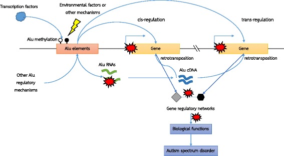
Background: Alu elements are a group of repetitive elements that can influence gene expression through CpG residues and transcription factor binding. Altered gene expression and methylation profiles have been reported in various tissues and cell lines from individuals with autism spectrum disorder (ASD). However, the role of Alu elements in ASD remains unclear. We thus investigated whether Alu elements are associated with altered gene expression profiles in ASD.

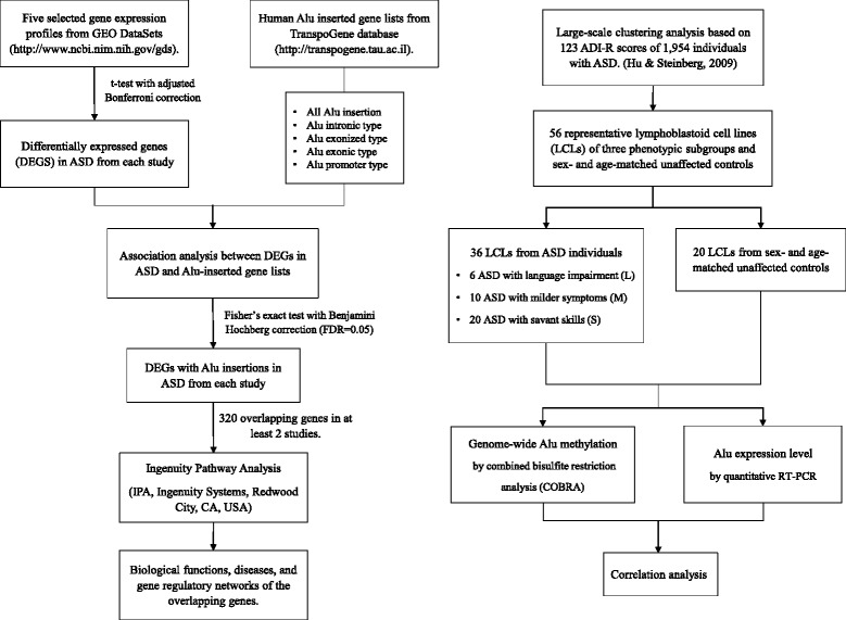
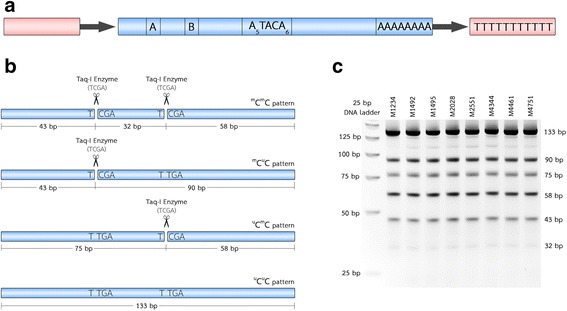
Methods: We obtained five blood-based gene expression profiles from the Gene Expression Omnibus database and human Alu-inserted gene lists from the TranspoGene database. Differentially expressed genes (DEGs) in ASD were identified from each study and overlapped with the human Alu-inserted genes. The biological functions and networks of Alu-inserted DEGs were then predicted by Ingenuity Pathway Analysis (IPA). A combined bisulfite restriction analysis of lymphoblastoid cell lines (LCLs) derived from 36 ASD and 20 sex- and age-matched unaffected individuals was performed to assess the global DNA methylation levels within Alu elements, and the Alu expression levels were determined by quantitative RT-PCR.

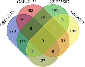
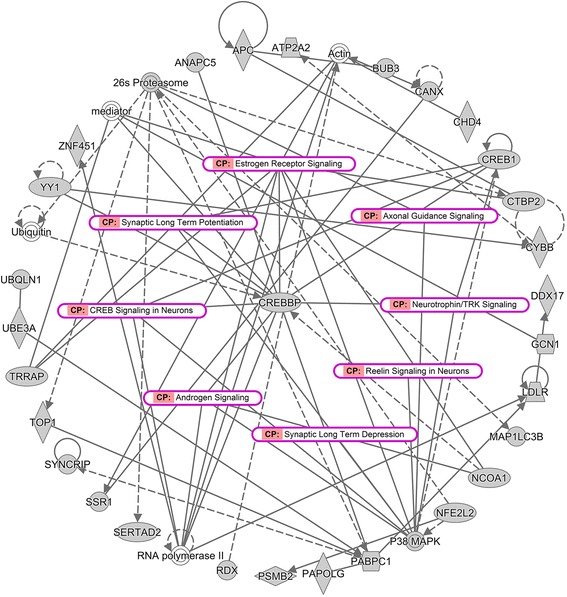
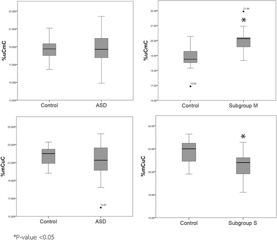
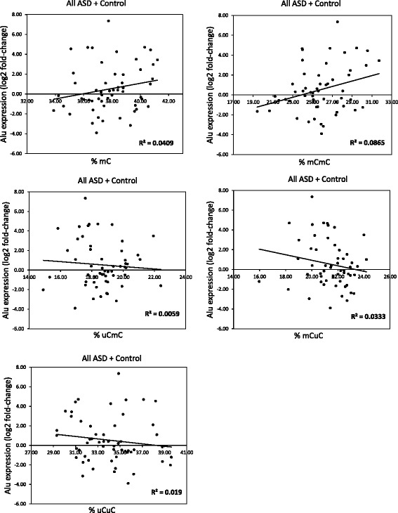
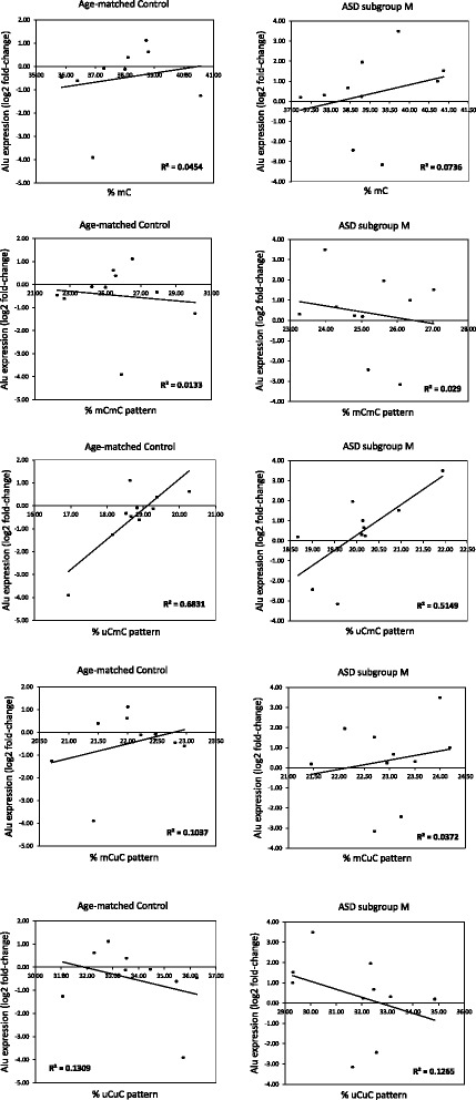
Results: In ASD blood or blood-derived cells, 320 Alu-inserted genes were reproducibly differentially expressed. Biological function and pathway analysis showed that these genes were significantly associated with neurodevelopmental disorders and neurological functions involved in ASD etiology. Interestingly, estrogen receptor and androgen signaling pathways implicated in the sex bias of ASD, as well as IL-6 signaling and neuroinflammation signaling pathways, were also highlighted. Alu methylation was not significantly different between the ASD and sex- and age-matched control groups. However, significantly altered Alu methylation patterns were observed in ASD cases sub-grouped based on Autism Diagnostic Interview-Revised scores compared with matched controls. Quantitative RT-PCR analysis of Alu expression also showed significant differences between ASD subgroups. Interestingly, Alu expression was correlated with methylation status in one phenotypic ASD subgroup.

Conclusion: Alu methylation and expression were altered in LCLs from ASD subgroups. Our findings highlight the association of Alu elements with gene dysregulation in ASD blood samples and warrant further investigation. Moreover, the classification of ASD individuals into subgroups based on phenotypes may be beneficial and could provide insights into the still unknown etiology and the underlying mechanisms of ASD.

Keywords: Alu elements; Autism spectrum disorder; DNA methylation; Epigenetic regulation; Gene expression profiles; Lymphoblastoid cell lines; Neuroinflammation; Retrotransposon; Sex bias; Subgrouping.


Conflict of interest statement

Not applicable.The authors declare that they have no competing interests.Springer Nature remains neutral with regard to jurisdictional claims in published maps and institutional affiliations.

Figures

Fig. 1
Fig. 1
Schematic diagram of experimental workflow. Our workflow initiated with the acquisition of blood-based gene expression profiles from GEO DataSets and human Alu-inserted gene lists. Fisher’s exact test was then used to identify differentially expressed genes (DEGs) with Alu insertions. A total of 320 overlapping genes among the selected study results were used to predict biological functions, diseases, and gene regulatory networks. Fifty-six LCLs were used as a model to investigate the association between the Alu methylation status and Alu expression profiles in LCLs
Fig. 2
Fig. 2
Alu element structure and illustration of COBRA for determining AluS methylation levels and patterns. a Alu elements are approximately 300 bp in length and have a dimeric structure that is separated by an A-rich region (A5TACA6) and ends with a poly-A tail. The left half of the Alu contains the A and B boxes, which are internal promoters for RNA polymerase III. b Illustration of the COBRA method designed to assess methylation of two CpGs at the internal promoter of AluS subfamilies. The four different methylation patterns of AluS were calculated from the percentages of differently digested products of 133, 75, 58, 43, and 32 bp. c Representative gel image from the COBRA for AluS subfamilies
Fig. 3
Fig. 3
Venn diagram of genes containing Alu that are differentially expressed in ASD. The significant DEGs with Alu insertions from each study based on Fisher’s exact test overlapped. The diagram shows the reproducibility of Alu-inserted genes that were differentially expressed in peripheral blood and blood-derived cell lines from ASD individuals. A total of 320 genes were selected to identify biological functions and gene regulatory networks through an Ingenuity Pathway Analysis (IPA)
Fig. 4
Fig. 4
Predicted gene regulatory network of the overlapping genes associated with neurological disease. This network revealed interactions or relationships among the overlapping molecules (gray background) and with other molecules from the IPA database (white background) that play a role in several mechanisms associated with neurological disease and estrogen receptor and androgen signaling, which is known to be associated with sex bias in ASD (labeled pink)
Fig. 5
Fig. 5
Box plot of the Alu methylation patterns in the LCLs of ASD subgroup M. In ASD subgroup M, the percentage of the partially methylated pattern uCmC (20.06% ± 0.92%) was significantly increased. In ASD subgroup S, the partially methylated pattern mCuC was significantly decreased. *adjusted P value < 0.05
Fig. 6
Fig. 6
Correlation analysis between AluS methylation and expression level for all LCL samples. The AluS expression for each LCL was normalized with the average GAPDH dCt of the control group. The Alu expression levels were then calculated using the 2−ΔΔCt method
Fig. 7
Fig. 7
Correlation analysis between AluS methylation and expression levels in ASD subgroup M and sex- and age-matched controls. The AluS expression of each LCL was normalized to the average GAPDH dCt of the control group. The Alu expression levels were then calculated using the 2−ΔΔCt method
Fig. 8
Fig. 8
Schematic diagram illustrating a possible mechanism of Alu elements in ASD. Our model suggests that exposure to environmental factors or dysregulation of other DNA methylation regulatory mechanisms lead to changes in CpG methylation patterns in Alu elements. Such changes alter transcription factor binding and, possibly in combination with other Alu regulatory mechanisms, cause the dysregulation of the expression and retrotransposition of Alu elements. Disrupted Alu retrotransposition results in changes in target genes via cis-/trans-regulatory mechanisms, which, in turn, dysregulate gene expression and gene regulatory networks known to be negatively impacted in ASD

References

    1. American Psychiatric Association: Diagnostic and Statistical Manual of Mental Disorders. 5th edition. Arlington, VA: 2013.
    1. Christensen DL, Baio J, Braun KV, Bilder D, Charles J, Constantino JN, Daniels J, Durkin MS, Fitzgerald RT, Kurzius-Spencer M, et al. Prevalence and characteristics of autism spectrum disorder among children aged 8 years—autism and developmental disabilities monitoring network, 11 sites, United States, 2012. Morbidity Mortal Weekl Repo Surveillance Summ (Washington, DC : 2002) 2016;65(3):1–23. - PMC - PubMed
    1. Lavelle TA, Weinstein MC, Newhouse JP, Munir K, Kuhlthau KA, Prosser LA. Economic burden of childhood autism spectrum disorders. Pediatrics. 2014;133(3):e520–e529. doi: 10.1542/peds.2013-0763. - DOI - PMC - PubMed
    1. Schaefer GB, Mendelsohn NJ. Genetics evaluation for the etiologic diagnosis of autism spectrum disorders. Genet Med. 2008;10(1):4–12. doi: 10.1097/GIM.0b013e31815efdd7. - DOI - PubMed
    1. Moosa A, Shu H, Sarachana T, Hu VW. Are endocrine disrupting compounds environmental risk factors for autism spectrum disorder? Horm Behav. 2017. 10.1016/j.yhbeh.2017.10.003. - PMC - PubMed

Publication types