

Head and Neck Schwannomas: A Surgical Challenge-A Series of 5 Cases
- PMID: 29686918
- PMCID: PMC5857344
- DOI: 10.1155/2018/4074905
Head and Neck Schwannomas: A Surgical Challenge-A Series of 5 Cases
Abstract
Background: Schwannomas, also known as neurilemmomas, are benign peripheral nerve sheath tumors. They originate from any nerve covered with schwann cell sheath. Schwannomas constitute 25-45% of tumors of the head and neck. About 4% of head and neck schwannomas present as a sinonasal schwannoma. Brachial plexus schwannoma constitute only about 5% of schwannomas. Cervical vagal schwannomas constitute about 2-5% of neurogenic tumors.
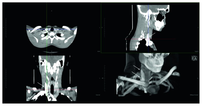
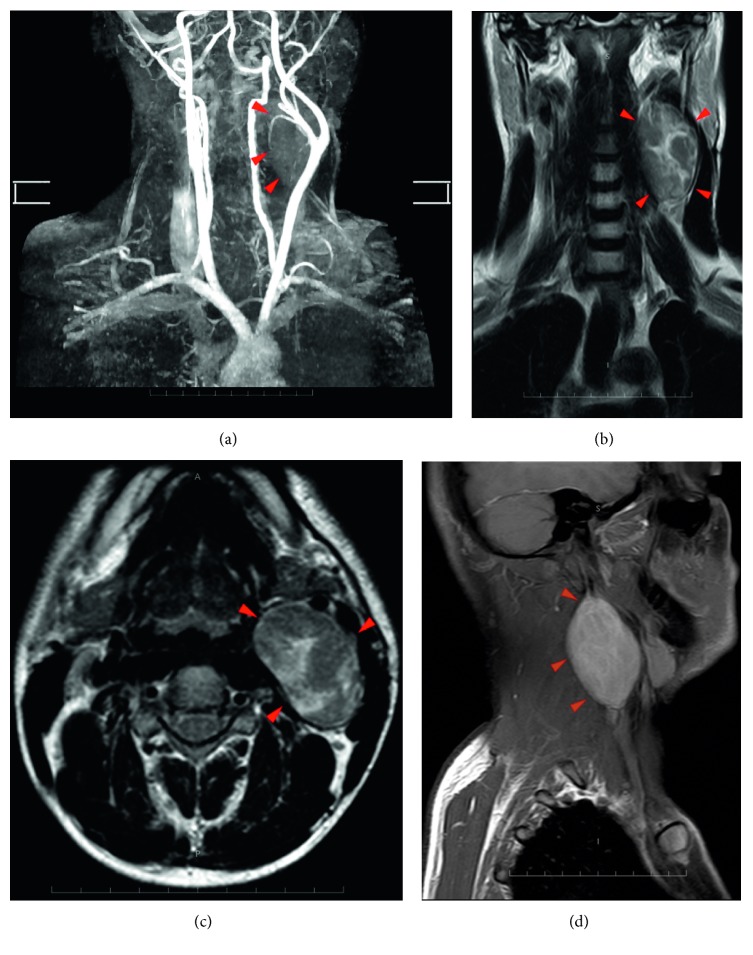
Methods: We present a case series of 5 patients of schwannomas, one arising from the maxillary branch of trigeminal nerve in the maxillary sinus, second arising from the brachial plexus, third arising from the cervical vagus, and two arising from cervical spinal nerves.
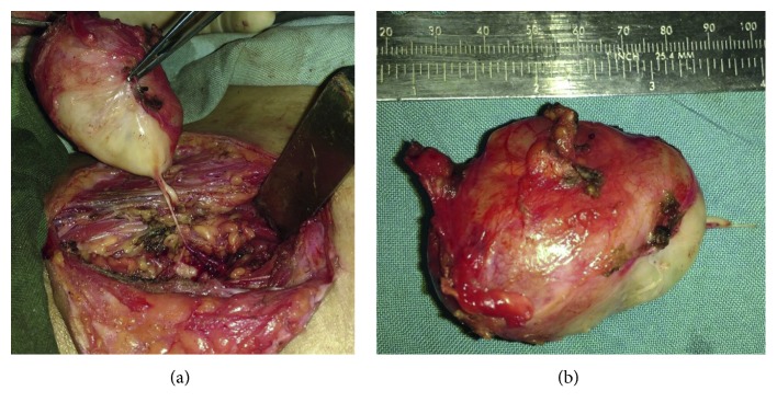
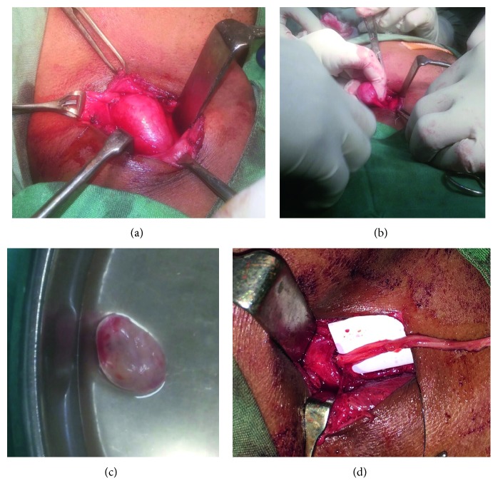
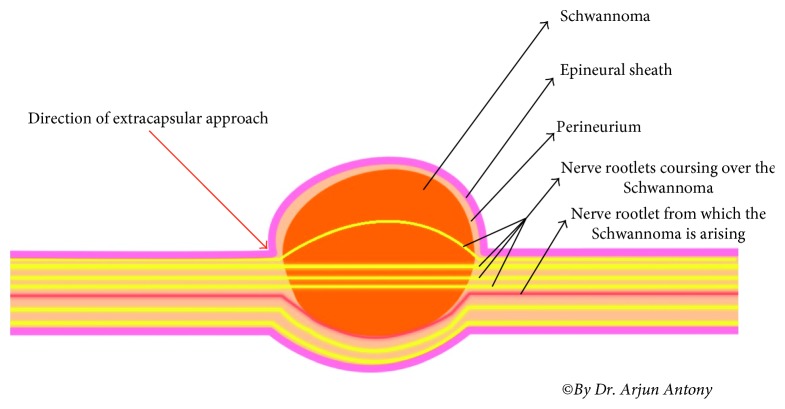
Result: Complete extracapsular excision of the tumors was achieved by microneurosurgical technique with preservation of nerve of origin in all except one.
Conclusion: Head and neck schwannoma though rare should be considered as a differential diagnosis of a unilateral slow growing mass in the head and neck region, particularly in an adult. Schwannomas are always a diagnostic dilemma as they are asymptomatic for long time, and histopathology is the gold standard for diagnosis. As a rule, treatment is surgical and dictated by the location of the tumor and nerve of origin. Due to its rarity, complex anatomical location and morbidity risk postexcision, they can pose a formidable challenge to surgeons. This study aims to describe the presentation, workup, surgical technique, and outcome.
Figures
Similar articles
-
A Rare Case of Cervical Vagal Nerve Schwannoma in a 30-Year-Old Ethiopian Man.Int Med Case Rep J. 2023 Mar 11;16:141-151. doi: 10.2147/IMCRJ.S401858. eCollection 2023. Int Med Case Rep J. 2023. PMID: 36936185 Free PMC article.
-
Extracranial non-vestibular head and neck schwannomas: a case series with the review of literature.Braz J Otorhinolaryngol. 2022 Nov-Dec;88 Suppl 4(Suppl 4):S9-S17. doi: 10.1016/j.bjorl.2021.05.013. Epub 2021 Jun 9. Braz J Otorhinolaryngol. 2022. PMID: 34154970 Free PMC article. Review.
-
Extracranial Head and Neck Schwannomas: Our Experience.Indian J Otolaryngol Head Neck Surg. 2016 Jun;68(2):241-7. doi: 10.1007/s12070-015-0925-5. Epub 2015 Dec 16. Indian J Otolaryngol Head Neck Surg. 2016. PMID: 27340644 Free PMC article.
-
Sinonasal and Infratemporal Schwannoma: Rare Case Report with Literature Review.Indian J Otolaryngol Head Neck Surg. 2023 Apr;75(Suppl 1):234-241. doi: 10.1007/s12070-022-03424-3. Epub 2022 Dec 21. Indian J Otolaryngol Head Neck Surg. 2023. PMID: 37206829 Free PMC article.
-
Extracranial Schwannomas of the Head and Neck: A Literature Review and Audit of Diagnosed Cases Over a Period of Eight Years.Head Neck Pathol. 2022 Sep;16(3):707-715. doi: 10.1007/s12105-022-01415-y. Epub 2022 Feb 14. Head Neck Pathol. 2022. PMID: 35157211 Free PMC article. Review.
Cited by
-
Case Report: Hemodynamic challenges during the anesthetic management of a patient who presented with a cervical vagal schwannoma in southern Ethiopia: a rare clinical case.Front Surg. 2025 Jun 24;12:1569722. doi: 10.3389/fsurg.2025.1569722. eCollection 2025. Front Surg. 2025. PMID: 40630866 Free PMC article.
-
Cervical vagal nerve schwannoma induced arrhythmia: a rare case report and literature review.BMC Neurol. 2022 Dec 14;22(1):480. doi: 10.1186/s12883-022-03016-2. BMC Neurol. 2022. PMID: 36517768 Free PMC article. Review.
-
Intracapsular Micro-Enucleation of a Painful Superficial Peroneal Nerve Schwannoma in a 60-Year Old Man: A Rare Encounter.Am J Case Rep. 2022 Feb 24;23:e936056. doi: 10.12659/AJCR.936056. Am J Case Rep. 2022. PMID: 35197439 Free PMC article.
-
Cervical Schwannoma: Diagnosis and Treatment in a Second-Level Hospital in Mexico.Cureus. 2025 Jul 7;17(7):e87481. doi: 10.7759/cureus.87481. eCollection 2025 Jul. Cureus. 2025. PMID: 40777668 Free PMC article.
-
Trigeminocardiac Reflex as a Complication of Excision of Schwannoma of the Trigeminal Nerve - A Rare Clinical Case Report.Ann Maxillofac Surg. 2022 Jul-Dec;12(2):216-218. doi: 10.4103/ams.ams_141_20. Epub 2023 Jan 10. Ann Maxillofac Surg. 2022. PMID: 36874786 Free PMC article.
References
-
- Batsakis J. G. Tumours of the Peripheral Nervous System. 2nd. Baltimore, MD, USA: Williams and Wilkins; 1979.
Publication types
LinkOut - more resources
Full Text Sources
Other Literature Sources
Research Materials
