. 2018 Jun;23(1-2):59-73.
doi: 10.1007/s10911-018-9392-4. Epub 2018 Apr 23.

Psychosocial Stress Exposure Disrupts Mammary Gland Development

Affiliations

Psychosocial Stress Exposure Disrupts Mammary Gland Development

Marianna B Johnson et al. J Mammary Gland Biol Neoplasia. 2018 Jun.

Abstract

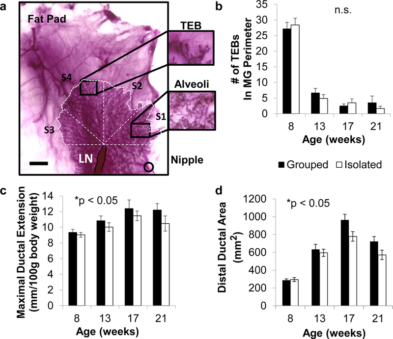
Exposure to psychosocial stressors and ensuing stress physiology have been associated with spontaneous invasive mammary tumors in the Sprague-Dawley rat model of human breast cancer. Mammary gland (MG) development is a time when physiologic and environmental exposures influence breast cancer risk. However, the effect of psychosocial stress exposure on MG development remains unknown. Here, in the first comprehensive longitudinal study of MG development in nulliparous female rats (from puberty through young adulthood; 8-25 wks of age), we quantify the spatial gradient of differentiation within the MG of socially stressed (isolated) and control (grouped) rats. We then demonstrate that social isolation increased stress reactivity to everyday stressors, resulting in downregulation of glucocorticoid receptor (GR) expression in the MG epithelium. Surprisingly, given that chemical carcinogens increase MG cancer risk by preventing normal terminal end bud (TEB) differentiation, chronic isolation stress did not alter TEBs. Instead, isolation blunted MG growth and alveolobular differentiation and reduced epithelial cell proliferation in these structures. Social isolation also enhanced corpora luteal progesterone at all ages but reduced estrogenization only in early adulthood, a pattern that precludes modulated ovarian function as a sufficient mechanism for the effects of isolation on MG development. This longitudinal study of natural variation provides an integrated view of MG development and the importance of increased GR activation in nulliparous ductal growth and alveolobular differentiation. Thus, social isolation and its physiological sequelae disrupt MG growth and differentiation and suggest a contribution of stress exposure during puberty and young adulthood to the previously observed increase in invasive MG cancer observed in chronically socially-isolated adult Sprague-Dawley rats.

Keywords: Alveolobular development; Mammary gland; Puberty; Stress; Terminal end buds; Young adulthood.


Conflict of interest statement

The authors declare that they have no conflict of interest.

Figures

Fig. 1
Fig. 1. Effect of social environment on mammary gland (MG) development of terminal end buds, ductal extension, and distal ductal area (measured in MG whole mounts).
a, a representative digital image of a MG from a group-housed Sprague-Dawley rat (8 weeks of age) stained with carmine alum, indicating the fat pad, lymph node (LN) terminal end buds (TEBs), and nipple. Scale bar = 5 mm. b, c, d. Effects of social condition on MG structures from puberty to early adulthood (8, 13, 17, and 21 weeks of age), measuring (b) TEB number at the periphery, (c) maximal ductal extension, and (d) distal ductal area; mean ± SEM; ≥ nine rats per age and social condition. *P < 0.05, 3-factor ANOVA (housing, age, and cohort).
Fig. 2
Fig. 2. Differentiation of ductal structures in four different sectors of the distal rat mammary gland (MG) (S1 nearest the nipple to S4 most distal).
A representative digital image of a MG from a group-housed Sprague-Dawley rat (8 weeks of age) stained with carmine alum. White dashed lines depict maximal ductal extension, the perimeter and four equal sectors (S1-S4) within the distal ductal area. Scale bar = 5 mm. TEB (terminal end bud), TD (terminal duct), AB (alveolar bud), L (lobule). The most prevalent structure is listed first, followed by the secondary structure: Individual variation is indicated by the % of rats at each age that had a specific pattern of primary and secondary structures. The spatial gradient in MG differentiation was greater than differences across chronological age.
Fig. 3
Fig. 3. Characterization of mammary gland (MG) composition.
Representative H&E stained cross-sections rostral to the lymph node of the left inguinal MG, at 17, 21, and 25 weeks of age (a) Ductal Structures (D), fat (F), and stroma (S), 5X magnification, scale bar = 500 µm and (b) 20X magnification, scale bar = 100 µm. c, Effect of social condition on % MG area occupied by ducts, stroma, and fat (mean ± SEM, ≥ four rats per age and social condition; 2-factor ANOVA (social condition and age), * interaction P < 0.05).
Fig. 4
Fig. 4. Epithelial cell proliferation in ducts, alveolar buds (ABs) and lobules (L) of the mammary gland (MG).
(a) Ki67 staining (brown) in representative cross-sections rostral to the lymph node of the left inguinal MG in early adulthood (21–25 weeks of age). Magnification 10X, scale bar = 200um. (b) Effect of social condition on %Ki67 in three mammary structures: ducts, ABs and Ls; 3-factor repeated measures ANOVA (social condition x structure interaction P = 0.02).
Fig. 5
Fig. 5. Vigilance testing, glucocorticoid stress reactivity, and adrenal weights.
a, percentage of rats emerging from home base in an open-field test (13–25 weeks of age). Log rank Mantle-Cox test, ***P < 0.001 b, corticosterone stress reactivity and recovery during and after a passive restraint stressor (Lined bar; 17 weeks; 30 minutes post restraint *P < 0.05 Student’s t-test). c, total adrenal gland weight (mg/100g body weight; mean ± SEM. **P <0.01, 3-factor ANOVA (housing, age, and cohort).
Fig. 6
Fig. 6. Ovarian steroid exposure (measured by vaginal cytology and ovarian weight).
a, number of days of luteal progesterone exposure per 14-day epoch. b, number of days of high estrogenization per 14-day epoch. c, average ovarian weight (mg/100g body weight). Mean ± SEM; ≥ nine rats per age and social condition. *P < 0.02, ***P < 0.001 3-factor ANOVA (housing, age, and cohort).
Fig. 7
Fig. 7. Characterization estrogen, progesterone, and glucocorticoid receptors.
Representative cross-sections rostral to the lymph node of the left inguinal MG at 13, 17, 21, and 25 weeks of age stained for (a) estrogen receptor (red), (b) progesterone receptor (brown), and (c) glucocorticoid receptor (brown). Arrows indicate positive receptor expression staining; for a-c, 20X magnification, scale bar = 100 µm. d, high resolution images of each receptor (ER, PR, and GR) expression at 21 weeks of age, scale bar = 50 µm. For all slides a-d, Hematoxylin (blue) was used for a counterstain.

References

    1. Hermes GL, Delgado B, Tretiakova M, Cavigelli SA, Krausz T, Conzen SD et al. Social isolation dysregulates endocrine and behavioral stress while increasing malignant burden of spontaneous mammary tumors. Proc Natl Acad Sci U S A 2009;106(52):22393–8. doi:10.1073/pnas.0910753106. - DOI - PMC - PubMed
    1. Russo J, Mailo D, Hu YF, Balogh G, Sheriff F, Russo IH. Breast differentiation and its implication in cancer prevention. Clin Cancer Res 2005;11(2 Pt 2):931s-6s. - PubMed
    1. MacMahon B, Cole P, Lin TM, Lowe CR, Mirra AP, Ravnihar B et al. Age at first birth and breast cancer risk. Bull World Health Organ 1970;43(2):209–21. - PMC - PubMed
    1. Colditz GA, Frazier AL. Models of breast cancer show that risk is set by events of early life: prevention efforts must shift focus. Cancer Epidemiol Biomarkers Prev 1995;4(5):567–71. - PubMed
    1. Osborne G, Rudel R, Schwarzman M. Evaluating chemical effects on mammary gland development: A critical need in disease prevention . Reprod Toxicol 2015;54:148–55. doi:10.1016/j.reprotox.2014.07.077. - DOI - PubMed

Publication types

LinkOut - more resources