

Amygdala real-time functional magnetic resonance imaging neurofeedback for major depressive disorder: A review
- PMID: 29687527
- PMCID: PMC6035103
- DOI: 10.1111/pcn.12665
Amygdala real-time functional magnetic resonance imaging neurofeedback for major depressive disorder: A review
Abstract
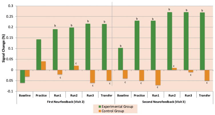
Advances in imaging technologies have allowed for the analysis of functional magnetic resonance imaging data in real-time (rtfMRI), leading to the development of neurofeedback (nf) training. This rtfMRI-nf training utilizes functional magnetic resonance imaging (fMRI) tomographic localization capacity to allow a person to see and regulate the localized hemodynamic signal from his or her own brain. In this review, we summarize the results of several studies that have developed and applied neurofeedback training to healthy and depressed individuals with the amygdala as the neurofeedback target and the goal to increase the hemodynamic response during positive autobiographical memory recall. We review these studies and highlight some of the challenges and advances in developing an rtfMRI-nf paradigm for broader use in psychiatric populations. The work described focuses on our line of research aiming to develop the rtfMRI-nf into an intervention, and includes a discussion of the selection of a region of interest for feedback, selecting a control condition, behavioral and cognitive effects of training, and predicting which participants are most likely to respond well to training. While the results of these studies are encouraging and suggest the clinical potential of amygdala rtfMRI-nf in alleviating symptoms of major depressive disorder, larger studies are warranted to confirm its efficacy.
Keywords: amygdala; autobiographical memory; emotional processing; functional magnetic resonance imaging neurofeedback; major depressive disorder.
© 2018 The Author. Psychiatry and Clinical Neurosciences © 2018 Japanese Society of Psychiatry and Neurology.
Figures
Similar articles
-
Randomized Clinical Trial of Real-Time fMRI Amygdala Neurofeedback for Major Depressive Disorder: Effects on Symptoms and Autobiographical Memory Recall.Am J Psychiatry. 2017 Aug 1;174(8):748-755. doi: 10.1176/appi.ajp.2017.16060637. Epub 2017 Apr 14. Am J Psychiatry. 2017. PMID: 28407727 Free PMC article. Clinical Trial.
-
Altered task-based and resting-state amygdala functional connectivity following real-time fMRI amygdala neurofeedback training in major depressive disorder.Neuroimage Clin. 2017 Dec 5;17:691-703. doi: 10.1016/j.nicl.2017.12.004. eCollection 2018. Neuroimage Clin. 2017. PMID: 29270356 Free PMC article.
-
Real-Time Functional Magnetic Resonance Imaging Amygdala Neurofeedback Changes Positive Information Processing in Major Depressive Disorder.Biol Psychiatry. 2017 Oct 15;82(8):578-586. doi: 10.1016/j.biopsych.2017.03.013. Epub 2017 Mar 28. Biol Psychiatry. 2017. PMID: 28476207 Free PMC article. Clinical Trial.
-
Quality and denoising in real-time functional magnetic resonance imaging neurofeedback: A methods review.Hum Brain Mapp. 2020 Aug 15;41(12):3439-3467. doi: 10.1002/hbm.25010. Epub 2020 Apr 25. Hum Brain Mapp. 2020. PMID: 32333624 Free PMC article. Review.
-
Amygdala Modulation During Emotion Regulation Training With fMRI-Based Neurofeedback.Front Hum Neurosci. 2019 Mar 26;13:89. doi: 10.3389/fnhum.2019.00089. eCollection 2019. Front Hum Neurosci. 2019. PMID: 30971906 Free PMC article.
Cited by
-
The power of negative and positive episodic memories.Cogn Affect Behav Neurosci. 2022 Oct;22(5):869-903. doi: 10.3758/s13415-022-01013-z. Epub 2022 Jun 14. Cogn Affect Behav Neurosci. 2022. PMID: 35701665 Free PMC article. Review.
-
Modulating the interhemispheric activity balance in the intraparietal sulcus using real-time fMRI neurofeedback: Development and proof-of-concept.Neuroimage Clin. 2020;28:102513. doi: 10.1016/j.nicl.2020.102513. Epub 2020 Nov 27. Neuroimage Clin. 2020. PMID: 33396000 Free PMC article.
-
Altered spontaneous brain activity in patients with diabetic optic neuropathy: A resting-state functional magnetic resonance imaging study using regional homogeneity.World J Diabetes. 2021 Mar 15;12(3):278-291. doi: 10.4239/wjd.v12.i3.278. World J Diabetes. 2021. PMID: 33758647 Free PMC article. Clinical Trial.
-
Examination of the importance of anger/irritability and limited prosocial emotion/callous-unemotional traits to understand externalizing symptoms and adjustment problems in adolescence: A 10-year longitudinal study.Front Psychiatry. 2022 Sep 29;13:939603. doi: 10.3389/fpsyt.2022.939603. eCollection 2022. Front Psychiatry. 2022. PMID: 36245864 Free PMC article.
-
Activation of endogenous retrovirus triggers microglial immuno-inflammation and contributes to negative emotional behaviors in mice with chronic stress.J Neuroinflammation. 2023 Feb 15;20(1):37. doi: 10.1186/s12974-023-02724-x. J Neuroinflammation. 2023. PMID: 36793064 Free PMC article.
References
-
- Cox RW, Jesmanowicz A, Hyde JS. Real-time functional magnetic resonance imaging. Magn Reson Med. 1995;33:230–236. - PubMed
-
- deCharms RC. Applications of real-time fMRI. Nat Rev Neurosci. 2008;9:720–729. - PubMed
-
- deCharms RC, Christoff K, Glover GH, Pauly JM, Whitfield S, Gabrieli JD. Learned regulation of spatially localized brain activation using real-time fMRI. Neuroimage. 2004;21:436–443. - PubMed
-
- Weiskopf N, Sitaram R, Josephs O, Veit R, Scharnowski F, Goebel R, Birbaumer N, Deichmann R, Mathiak K. Real-time functional magnetic resonance imaging: methods and applications. Magn Reson Imaging. 2007;25:989–1003. - PubMed
-
- Yoo SS, O’Leary HM, Fairneny T, Chen NK, Panych LP, Park H, Jolesz FA. Increasing cortical activity in auditory areas through neurofeedback functional magnetic resonance imaging. Neuroreport. 2006;17:1273–1278. - PubMed
Publication types
MeSH terms
Grants and funding
LinkOut - more resources
Full Text Sources
Other Literature Sources
Medical
