

doi: 10.1093/database/bay002.
Worldwide Protein Data Bank biocuration supporting open access to high-quality 3D structural biology data
Affiliations
- PMID: 29688351
- PMCID: PMC5804564
- DOI: 10.1093/database/bay002
Item in Clipboard
Worldwide Protein Data Bank biocuration supporting open access to high-quality 3D structural biology data
Database (Oxford).
.
Abstract
https://www.wwpdb.org/.
Figures

Semantic relationships in the PDBx/mmCIF dictionary. The partial diagram shows the relationships within an entity, its polymer sequence, source taxonomy and the method used to produce it. The relationships in these categories are described by a shared key identifier in a parent/child relationship as denoted with gray shading. The dictionary is available at mmcif.wwpdb.org/ .

Major modules controlled by the workflow and WFM. (A) Mandatory major tasks controlled by the workflow with a pre-defined order. (B) WFM interface that allows Biocurators to prioritize their tasks, to manage multiple entries and to access module user interface for manual biocuration after completion of automated calculations.
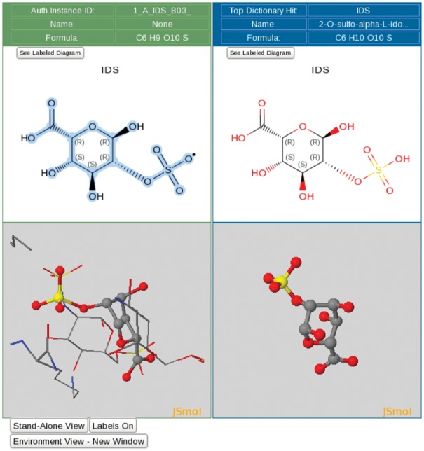
Ligand processing. The ligand processing module enables comparison of the structure of the deposited ligand with matches from the CCD. The top panels compare 2D structures, and the bottom panels compare 3D views of the model with matched ligands.

Examples of sequence alignments from the sequence processing module. (A) Alignment between the sample sequence (labeled ‘AUTH Entity’), the sequence extracted from the atom data for each polymer chain and the corresponding UniProt sequence. Sequence discrepancies are highlighted in red and listed in a table where the appropriate annotation can be selected. For example, residues 1–9 in the sample sequence (top sequence in the alignment) are not present in the UniProt sequence (bottom sequence in the alignment) because these are part of an expression tag. Similarly, the Tyrosine-Tryptophan (TYR-TRP) conflict at position 64 is annotated as a mutation based on information provided by the Depositor during data submission. (B) Example illustrating the sequence and 3D viewer. Residues depicted in orange in the sequence are highlighted and selected for visualization with the 3D viewer available on the alignment page. (C) Example of a sequence alignment for a chimeric protein construct. This chimeric acetylcholine-binding protein from Aplysia californica, PDB entry 5TVC, contains a loop C from the human alpha-3 nicotinic acetylcholine receptor. The alignment shows that residues 1–181 in the deposited sample sequence correspond to UniProt sequence Q8WSF8, residues 182–197 to UniProt sequence P32297 and residues 198–219 again to UniProt sequence Q8WSF8.

Comparison of ligand structures with 3D electron-density views. The electron-density maps shown in Figure 4A and B are 2m|Fo|-D|Fc| maps contoured at 1.0 σ cutoff. (A) Good electron-density fit for heparin oligosaccharide at residues 801–804 bound to annexin in PDB entry 2HYV. (B) Poor electron-density fit for NADP bound to alcohol dehydrogenase in PDB entry 1ZK4.

Average number of entries processed per wwPDB biocurator FTE and number of total global depositions per year. The processing productivity per wwPDB biocurator FTE has nearly doubled since 2008 as shown in this graph. This graph also reflects that the productivity was accelerated with the OneDep system. The label * indicates the transition period when both new OneDep and legacy systems were operated in parallel. The dash line indicates when the wwPDB validation report was first introduced in August 2013.
References
-
- Berman H.M., Henrick K., Nakamura H. (2003) Announcing the worldwide Protein Data Bank. Nat. Struct. Biol., 10, 980.. - PubMed
Publication types
MeSH terms
Grants and funding
- MR/L007835/1/MRC_/Medical Research Council/United Kingdom
- BB/M020428/1/BB_/Biotechnology and Biological Sciences Research Council/United Kingdom
- BB/M011674/1/BB_/Biotechnology and Biological Sciences Research Council/United Kingdom
- R01 GM109046/GM/NIGMS NIH HHS/United States
- BB/M013146/1/BB_/Biotechnology and Biological Sciences Research Council/United Kingdom
- 88944/WT_/Wellcome Trust/United Kingdom
- BB/G022577/1/BB_/Biotechnology and Biological Sciences Research Council/United Kingdom
- BB/M020347/1/BB_/Biotechnology and Biological Sciences Research Council/United Kingdom
- 75968/WT_/Wellcome Trust/United Kingdom
- BB/K020013/1/BB_/Biotechnology and Biological Sciences Research Council/United Kingdom
- BB/K016970/1/BB_/Biotechnology and Biological Sciences Research Council/United Kingdom
- 104948/WT_/Wellcome Trust/United Kingdom
LinkOut - more resources
Full Text Sources
Other Literature Sources
