

Serial Culture Is Critical for In Vitro Development of Parthenogenetic Embryos in the Golden Syrian Hamster
- PMID: 29688743
- PMCID: PMC6421986
- DOI: 10.1089/cell.2017.0070
Serial Culture Is Critical for In Vitro Development of Parthenogenetic Embryos in the Golden Syrian Hamster
Abstract
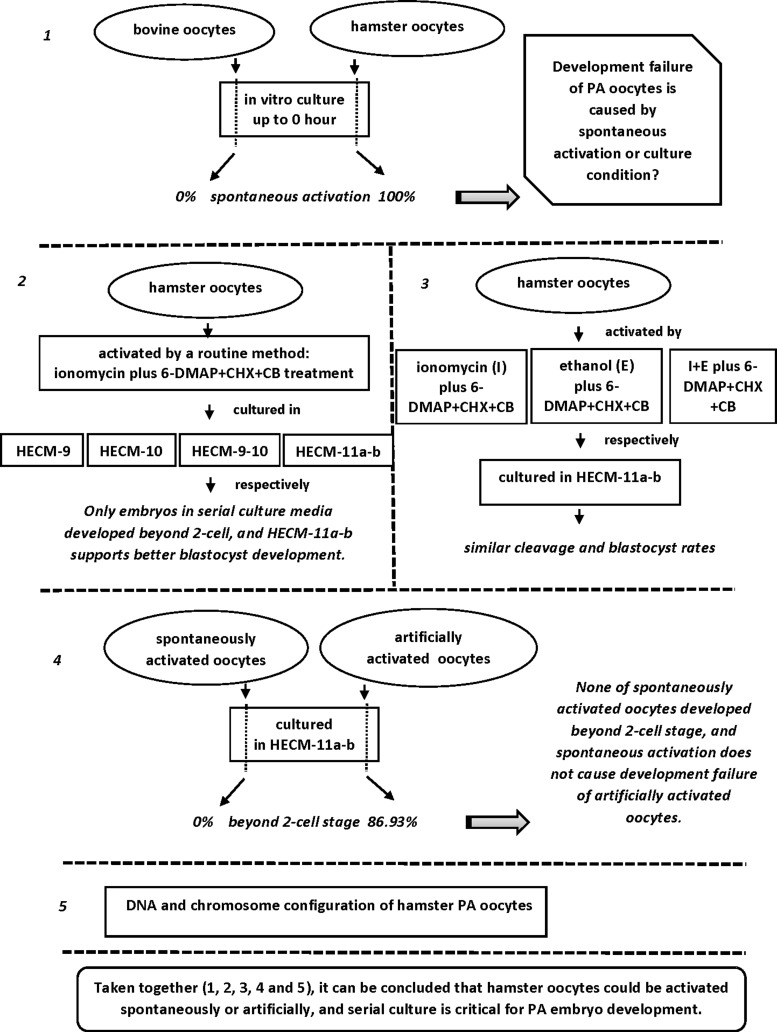
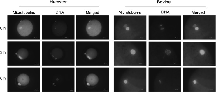
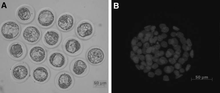
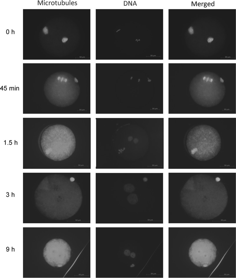
Unlike oocytes of many other mammalian species, parthenogenetically activated hamster oocytes have not been reported to develop beyond the two-cell stage. This study investigated the in vitro development into blastocysts of parthenogenetic embryos of Golden Syrian hamsters. We observed that hamster oocytes could easily be artificially activated (AA) by treatment with ionomycin plus 6-dimethylaminopurine + cycloheximide + cytochalasin B as assessed by embryo cleavage in HECM-9 (63.15%) or HECM-10 (63.82%). None of the cleaved embryos developed beyond the two-cell stage when cultured in either of the two media. However, some of the embryos overcame the two-cell block and developed to the blastocyst stage (26.45%) when they were first cultured in HECM-10 for 24 hours and then in HECM-9 (serial culture media HECM-10-9) for 72 hours. Blastocyst development was further significantly (66.2%) improved when embryos were cultured in HECM-10 supplemented with ethylenediaminetetraacetic acid for 24 hours, then in HECM-9 supplemented with glucose for 72 hours (serial culture media HECM-11a-b). Hamster oocytes activated with ionomycin, ethanol, or a combination of the two treatments would develop to the blastocyst stage in serial culture media HECM-11a-b, whereas none of the spontaneously activated oocytes cleaved (0% vs. 86.93%, p < 0.05). DNA and microtubule configurations of spontaneously activated and AA oocytes were assessed by immunocytochemical staining and fluorescence microscopy. The results indicate that serial culture and the method of activation are critical for overcoming the in vitro developmental block of hamster parthenogenetic embryos. This study is the first to report blastocyst development from parthenogenetically activated hamster oocytes.
Keywords: blastocyst; hamster oocytes; parthenogenetic activation; serial cultural media.
Conflict of interest statement
No competing financial interests exist.
Figures
References
-
- Balakier H., and Casper R.F. (1993). Experimentally induced parthenogenetic activation of human oocytes. Hum. Reprod. 8, 740–743 - PubMed
-
- Barros C., Gonzelez J., Herrera E., and Bustos-Obregon E. (1978). Fertilizing capacity of human spermatozoa evaluated by actual penetration of foreign eggs. Contraception 17, 87–92 - PubMed
-
- Bavister B.D., and Arlotto T. (1990). Influence of single amino acids on the development of hamster one-cell embryos in vitro. Mol. Reprod. Dev. 25, 45–51 - PubMed
-
- Chatot C.L., Lewis-Williams J., Torres I., and Ziomek C.A. (1994). One-minute exposure of 4-cell mouse embryos to glucose overcomes morula block in CZB medium. Mol. Reprod. Dev. 37, 407–412 - PubMed
-
- Chatot C.L., Lewis J.L., Torres I., and Ziomek C.A. (1990). Development of 1-cell embryos from different strains of mice in CZB medium. Biol. Reprod. 42, 432–440 - PubMed
Publication types
MeSH terms
Substances
Grants and funding
LinkOut - more resources
Full Text Sources
Other Literature Sources
