

Brain vascular heterogeneity: implications for disease pathogenesis and design of in vitro blood-brain barrier models
- PMID: 29688865
- PMCID: PMC5911972
- DOI: 10.1186/s12987-018-0097-2
Brain vascular heterogeneity: implications for disease pathogenesis and design of in vitro blood-brain barrier models
Abstract
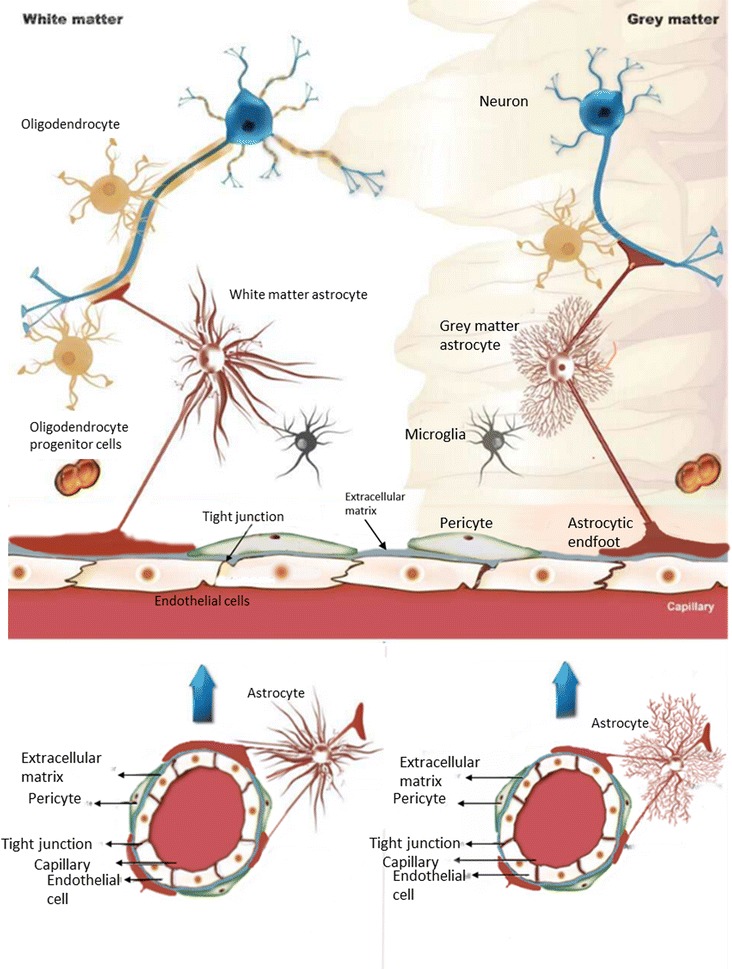
The vertebrate blood-brain barrier (BBB) is composed of cerebral microvascular endothelial cells (CEC). The BBB acts as a semi-permeable cellular interface that tightly regulates bidirectional molecular transport between blood and the brain parenchyma in order to maintain cerebral homeostasis. The CEC phenotype is regulated by a variety of factors, including cells in its immediate environment and within functional neurovascular units. The cellular composition of the brain parenchyma surrounding the CEC varies between different brain regions; this difference is clearly visible in grey versus white matter. In this review, we discuss evidence for the existence of brain vascular heterogeneity, focusing on differences between the vessels of the grey and white matter. The region-specific differences in the vasculature of the brain are reflective of specific functions of those particular brain areas. This BBB-endothelial heterogeneity may have implications for the course of pathogenesis of cerebrovascular diseases and neurological disorders involving vascular activation and dysfunction. This heterogeneity should be taken into account when developing BBB-neuro-disease models representative of specific brain areas.
Keywords: Blood–brain barrier; Cerebral vasculature; Grey matter; In vitro models; Neurodegeneration; Neurovascular unit; White matter.
Figures
References
-
- Abbott NJ. Blood–brain barrier structure and function and the challenges for CNS drug delivery. J Inherit Metabol Dis. 2013;36:437–449. - PubMed
-
- Abbott NJ, Patabendige AAK, Dolman DEM, Yusof SR, Begley DJ. Structure and function of the blood–brain barrier. Neurobiol Dis. 2010;37:13–25. - PubMed
-
- Wolburg H, Wolburg-Buchholz K, Engelhardt B. Diapedesis of mononuclear cells across cerebral venules during experimental autoimmune encephalomyelitis leaves tight junctions intact. Acta Neuropathol. 2005;109:181–190. - PubMed
-
- Lok J, Gupta P, Guo S, Kim WJ, Whalen MJ, van Leyen K, et al. Cell–cell signaling in the neurovascular unit. Neurochem Res. 2007;32:2032–2045. - PubMed
Publication types
MeSH terms
Grants and funding
LinkOut - more resources
Full Text Sources
Other Literature Sources
