

COMBINATION THERAPY OF INTRAVITREAL RANIBIZUMAB AND SUBTHRESHOLD MICROPULSE PHOTOCOAGULATION FOR MACULAR EDEMA SECONDARY TO BRANCH RETINAL VEIN OCCLUSION: 6-MONTH RESULT
- PMID: 29689025
- PMCID: PMC6613833
- DOI: 10.1097/IAE.0000000000002165
COMBINATION THERAPY OF INTRAVITREAL RANIBIZUMAB AND SUBTHRESHOLD MICROPULSE PHOTOCOAGULATION FOR MACULAR EDEMA SECONDARY TO BRANCH RETINAL VEIN OCCLUSION: 6-MONTH RESULT
Abstract
Purpose: To determine the efficacy of the combination therapy of intravitreal ranibizumab (IVR) and 577-nm yellow laser subthreshold micropulse laser photocoagulation (SMLP) for macular edema secondary to branch retinal vein occlusion cystoid macular edema.
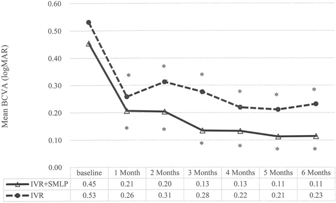
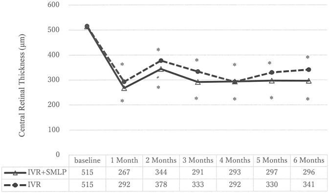
Methods: Retrospective, consecutive, case-control study. Forty-six eyes of 46 patients with treatment-naive branch retinal vein occlusion cystoid macular edema were enrolled. The IVR + SMLP group consisted of 22 patients who had undergone both SMLP and IVR. Intravitreal ranibizumab group consisted of 24 patients who had undergone IVR monotherapy. Intravitreal ranibizumab therapy was one initial injection and on a pro re nata in both groups, and SMLP was performed at 1 month after IVR in the IVR + SMLP group. Preoperatively and monthly, best-corrected visual acuity and central retinal thickness were evaluated using swept source optical coherence tomography.
Results: Best-corrected visual acuity and central retinal thickness significantly improved at 6 months in IVR + SMLP and IVR groups. Best-corrected visual acuity and central retinal thickness were not significantly different between the two groups at any time points. The number of IVR injections during initial 6 months in IVR group (2.3 ± 0.9) was significantly greater (P = 0.034) than that in IVR + SMLP group (1.9 ± 0.8).
Conclusion: The combination therapy of IVR and SMLP can treat branch retinal vein occlusion cystoid macular edema effectively, by decreasing the frequency of IVR injections while maintaining good visual acuity.
Conflict of interest statement
None of the authors has any financial/conflicting interests to disclose.
Figures
References
-
- Mitchell P, Smith W, Chang A. Prevalence and associations of retinal vein occlusion in Australia. The Blue Mountains Eye Study. Arch Ophthalmol 1996;114:1243–1247. - PubMed
-
- Glacet-Bernard A, Coscas G, Chabanel A, et al. Prognostic factors for retinal vein occlusion: prospective study of 175 cases. Ophthalmology 1996;103:551–560. - PubMed
-
- Finkelstein D. Ischemic macular edema. Recognition and favorable natural history in branch vein occlusion. Arch Ophthalmol 1992;110:1427–1434. - PubMed
-
- Wallow IH, Danis RP, Bindley C, Neider M. Cystoid macular degeneration in experimental branch retinal vein occlusion. Ophthalmology 1988;95:1371–1379. - PubMed
-
- Kreutzer TC, Alge CS, Wolf AH, et al. Intravitreal bevacizumab for the treatment of macular oedema secondary to branch retinal vein occlusion. Br J Ophthalmol 2008;92:351–355. - PubMed
MeSH terms
Substances
LinkOut - more resources
Full Text Sources
Other Literature Sources
