. 2018 Apr 24;13(4):e0195969.
doi: 10.1371/journal.pone.0195969. eCollection 2018.

Identification of RNA-binding proteins in exosomes capable of interacting with different types of RNA: RBP-facilitated transport of RNAs into exosomes

Affiliations

Identification of RNA-binding proteins in exosomes capable of interacting with different types of RNA: RBP-facilitated transport of RNAs into exosomes

Luisa Statello et al. PLoS One. .

Abstract

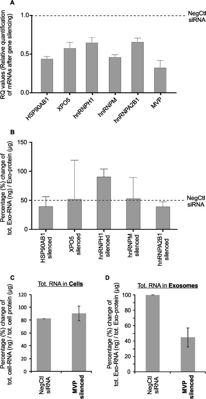
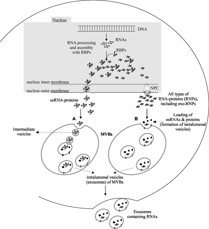
The RNA that is packaged into exosomes is termed as exosomal-shuttle RNA (esRNA); however, the players, which take this subset of RNA (esRNA) into exosomes, remain largely unknown. We hypothesized that RNA binding proteins (RBPs) could serve as key players in this mechanism, by making complexes with RNAs and transporting them into exosomes during the biosynthesis of exosomes. Here, we demonstrate the presence of 30 RBPs in exosomes that were shown to form RNA-RBP complexes with both cellular RNA and exosomal-RNA species. To assess the involvement of these RBPs in RNA-transfer into exosomes, the gene transcripts encoding six of the proteins identified in exosomes (HSP90AB1, XPO5, hnRNPH1, hnRNPM, hnRNPA2B1, and MVP) were silenced by siRNA and subsequent effect on esRNA was assessed. A significant reduction of total esRNA was observed by post-transcriptional silencing of MVP, compared to other RBPs. Furthermore, to confirm the binding of MVP with esRNA, a biotinylated-MVP was transiently expressed in HEK293F cells. Higher levels of esRNA were recovered from MVP that was eluted from exosomes of transfected cells, as compared to those of non-transfected cells. Our data indicate that these RBPs could end up in exosomes together with RNA molecules in the form of RNA-ribonucleoprotein complexes, which could be important for the transport of RNAs into exosomes and the maintenance of RNAs inside exosomes. This type of maintenance may favor the shuttling of RNAs from exosomes to recipient cells in the form of stable complexes.


Conflict of interest statement

Competing Interests: All the authors on this paper declare that the authors of this manuscript have no financial interest to this work. The following authors: A. Collén who is affiliated with AstraZeneca AB, Gothenburg, Sweden whereas, N. Tigue with MedImmune Ltd, Granta Park, Cambridge, UK declare no conflict of interests. This does not alter our adherence to PLOS ONE policies on sharing data and materials.

Figures

Fig 1
Fig 1
(A) RNA 3’-end labelling of cellular (miRNA, mRNA) and exosomal-RNAs (esRNA). The dot blot shows the efficiency of biotinylation of cellular miRNAs and mRNA, and exosomal total RNA (esRNA), relative to the biotinylated IRE control. A 43 nucleotide synthetic RNA template was used as a positive control for the reaction and as a general mass control. Serial dilutions of the labelled samples were loaded onto a nylon membrane, UV cross-linked, and detected by chemiluminescence. Dot blots show the efficiency of the RNA labelling reaction and the amount of the biotinylated RNA obtained. The biotinylation reaction for all RNA species esRNA, cell-miRNA and cell-mRNA was highly efficient, with biotinylation rates greater than 75%. (B) RNA electrophoretic mobility shift sssays (REMSAs) of cellular and exosomal RNA-RBP-complexes: Total exosomal-RNA (esRNA), cell-miRNA and cell-mRNA fractions extracted from HTB (lung) cells were incubated in binding reactions with exosomal proteins and with HTB cellular proteins. In all experiments, an electrophoretic shift was observed when the biotinylated RNA was incubated with the proteins from exosomes and cells. The specificities of the RNA-protein interactions were determined using competition assays. From left to right: the interaction of IRE RNA and cytosolic liver extract (positive control), the interaction of IRE RNA with cellular proteins, the interaction of cellular miRNAs with cellular proteins, the interaction of esRNA with exosomal proteins, and the interaction of cellular miRNA with exosomal proteins. The band-shifts are indicated by squares.
Fig 2
Fig 2. Identification of RNA-binding proteins (RBPs) from exosomes and their interactions with different RNA species.
Total exosomal proteins were isolated and incubated separately in independent assays; (total exosomal protein extract + esRNA; total exosomal protein extract + cell-mRNA; total exosomal protein extract + cell-miRNA) and their interaction with RNA species were identified. From total exosomal proteins, 30 were identified as RNA-binding proteins (i.e. exosomal-RBPs). (A) Schematic representation of biotinylation, streptavidin and LC-MS/MS experimental procedures used to label RNA, and to identify exosomal-RBPs respectively. (i) Total RNA isolated from exosomes, and cells from independent assays was biotinylated and streptavidin-coated Dynabeads were added. (ii) The isolated native total exosomal proteins were incubated separately with esRNA, cell-mRNA and cell-miRNA, and allowed to bind with biotinylated RNAs in separate reactions. (iii) Proteins were re-extracted and RBPs bound to the different RNAs were then loaded onto an SDS-PAGE gel, and target bands were excised, trypsinised and analyzed using LC-MS/MS. (B) List of the identified exosomal-RBPs from independent assays, which interact with cellular RNA and esRNA from different assays. (C) Venn diagrams displaying the overlap of 30 RBPs identified in exosomal-associated with exosomal RNA (esRNA), cellular mRNAs, and cellular miRNAs. In total, 30 different RBPs were identified in exosomes, including 20 in complex with esRNA. Out of 20 esRNA-binding exosomal-RBPs, the 12 exosomal-RBPs were exclusive to esRNA, 2 were common between the esRNA and cell-mRNA samples, 2 were common between the esRNA and cell-miRNA samples and 4 RBPs were common in all three samples i.e. esRNA, cell-mRNA and cell-miRNA. 9 exosomal-RBPs in complex with cell-miRNA, 14 in complex with cell-mRNA and 1 was common between cell-miRNA and cell-mRNA samples. 2 exosomal-RBPs were in complex with cell-miRNA only, and 7 were in complex with cell-mRNA only.
Fig 3
Fig 3. Network analysis of all (30) identified RBPs in exosomes.
The key function of the identified RBPs was retrieved using Ingenuity software (www.ingenuity.com) to build a biological network of the RBPs in exosomes. The network included 35 nodes (gene products), 27 of which were among the identified exosomal-RBPs including MVP (indicated in grey). The network shows the involvement of exosomal-RBPs in ‘RNA posttranscriptional modifications’.
Fig 4
Fig 4. The gene transcripts encoding six RBPs identified in exosomes were silenced in the cytoplasm by siRNA and the subsequent effect on the amount of esRNA was assessed.
A significant reduction of total RNA in exosomes (esRNA) was shown by post-transcriptional silencing of MVP. (A) Confirmation of gene silencing (silenced transcripts) in the cells after transfection with the siRNAs against the transcripts of HSP90AB1, XPO5, hnRNPH1, hnRNPM, hnRNPA2B1, and MVP with respect to the Negative Control siRNA (scrambled siRNA). Quantitative PCR was performed using cDNA, Fast SYBR Green Master Mix, and gene-specific primers. Gene expression was normalized to that of the housekeeping gene GAPDH. The error bars represent average of 4 biological replicates for hnRNPA2B1 and MVP, and 2 biological replicates for other four transcripts, and the average fold change values presented as RQ (relative quantification). (B) Quantification (the percentage-change, %) of total RNA present in exosomes (ng total esRNA / μg total exosomal proteins) after each gene silencing, i.e. silencing of HSP90AB1, XPO5, hnRNPH1, hnRNPM, and hnRNPA2B1. The silencing of hnRNPA2B1 caused a slight reduction of total RNA present in exosomes, but at non-significant level ≈13%. However, after silencing of hnRNPH1 the amount of RNA in exosomes was increased as compared to negative control. The experiment was performed in duplicates, except from hnRNPA2B1 that was performed in 4 replicates. The graph shows the range of values and median (grey). (C-D) Quantification of total RNA (ng total RNA / μg total protein) present in cells and exosomes after gene silencing of MVP. (C) A slight increase (but not significant, p-value 0.57) of the amount of total RNA in cytoplasm was observed. (D) On the contrary, down regulation of MVP (silencing) caused a significant reduction of the total RNA present in exosomes (esRNA), approximately by 50% (p-value 0.02). The experiment for MVP silencing was performed in four replicates; the graph shows the range of values and median (grey).
Fig 5
Fig 5. Validation of MVP.
Transfected cells are expressing MVP-biotin and un-transfected cells were used as negative control. (A) Western blot analysis for the detection of MVP in HEK293F cell lysate and exosomal protein extracts shows that plasmid (MVP-biotin) was successfully expressed in HEK293F cells and was partitioned into exosomes isolated from these transfected cells. (B) Protein samples from different stages of MVP purification were separated by SDS-PAGE and the protein bands were detected by Comassie staining. After the pull down assay, the presence of MVP within exosomes was confirmed, i.e. MVP in exosomes from biotinylated MVP-transfected cells PD (pull-down lane), whereas it was absent in exosomes from untransfected cells. The position of the MVP-biotin band is indicated by squares. A representative experiment out of 3 is shown. (C) Total amount of RNAs coupled to MVP were extracted and quantified by Quant-iT™ RiboGreen® RNA Assay Kit (ThermoFisher Scientific). Captured RNA was expressed as a percentage of RNA eluted from the beads after the pull-down respect to the total RNA incubated with the beads. Exosomal-MVP was coupled with RNAs and the quantification of RNAs that had co-eluted with MVP showed that the amount of RNA present in the MVP elute (i.e. from MVP-RNA complex) was significantly higher than that from the untransfected control. The experiment was performed in 3-replicates and the graph shows the range of values and median (grey). Average and standard error of three independent experiments are shown. Samples: W = proteins from different wash steps during the MVP purification, and PD = pull-down proteins (MVP) after elution, I = input, and UB = unbound after RBP capture.
Fig 6
Fig 6
Two hypothetical paths (A and B) for the packaging of RNA–protein–complexes (RNPs) into exosomes during biosynthesis of these vesicles from the multivesicular bodies (MVBs): (A) The RNP-granules i.e. RNA–protein complexes to be included into exosomes (so called esRNA-protein complex) use a budding mechanism to enter into intraluminal vesicles of MVBs as RNA-protein granules. In doing so, first, they form an intermediate vesicle inside the MVBs, by inward budding–a mechanism similar to nuclear envelop (NE) budding as shown by [–92]. The intermediate vesicles are matured and transformed into intraluminal vesicles inside MVB. Upon fusion of MVB with the plasma membrane, the intraluminal vesicles (exosomes) will be released outside containing RNA-RNPs complexes into the extracellular environment. In the second model (B) RNA–protein complexes (RNPs)—transported via nuclear pore complex (NPC)—are attracted to the internalization site having different receptor-proteins that could mediate inward budding of MVBs, to form intraluminal vesicles. Upon fusion with the plasma membrane, the MVBs release the intraluminal vesicles (exosomes) containing RNA–RNPs complexes into the extracellular environment.

References

    1. Fevrier B, Raposo G. Exosomes: endosomal-derived vesicles shipping extracellular messages. Current opinion in cell biology. 2004;16(4):415–21. doi: 10.1016/j.ceb.2004.06.003 - DOI - PubMed
    1. Nawaz M, Camussi G, Valadi H, Nazarenko I, Ekstrom K, Wang X, et al. The emerging role of extracellular vesicles as biomarkers for urogenital cancers. Nature reviews Urology. 2014;11(12):688–701. doi: 10.1038/nrurol.2014.301 - DOI - PubMed
    1. Fatima F, Nawaz M. Vesiculated Long Non-Coding RNAs: Offshore Packages Deciphering Trans-Regulation between Cells, Cancer Progression and Resistance to Therapies. Non-Coding RNA. 2017;3(1):10. - PMC - PubMed
    1. Mathivanan S, Ji H, Simpson RJ. Exosomes: extracellular organelles important in intercellular communication. Journal of proteomics. 2010;73(10):1907–20. doi: 10.1016/j.jprot.2010.06.006 - DOI - PubMed
    1. Ratajczak J, Wysoczynski M, Hayek F, Janowska-Wieczorek A, Ratajczak MZ. Membrane-derived microvesicles: important and underappreciated mediators of cell-to-cell communication. Leukemia. 2006;20(9):1487–95. doi: 10.1038/sj.leu.2404296 - DOI - PubMed

Publication types