Effects of Somatosensory Impairment on Participation After Stroke
- PMID: 29689179
- PMCID: PMC5915232
- DOI: 10.5014/ajot.2018.025114
Effects of Somatosensory Impairment on Participation After Stroke
Abstract
Objective: Our objective was to determine the effect of loss of body sensation on activity participation in stroke survivors.
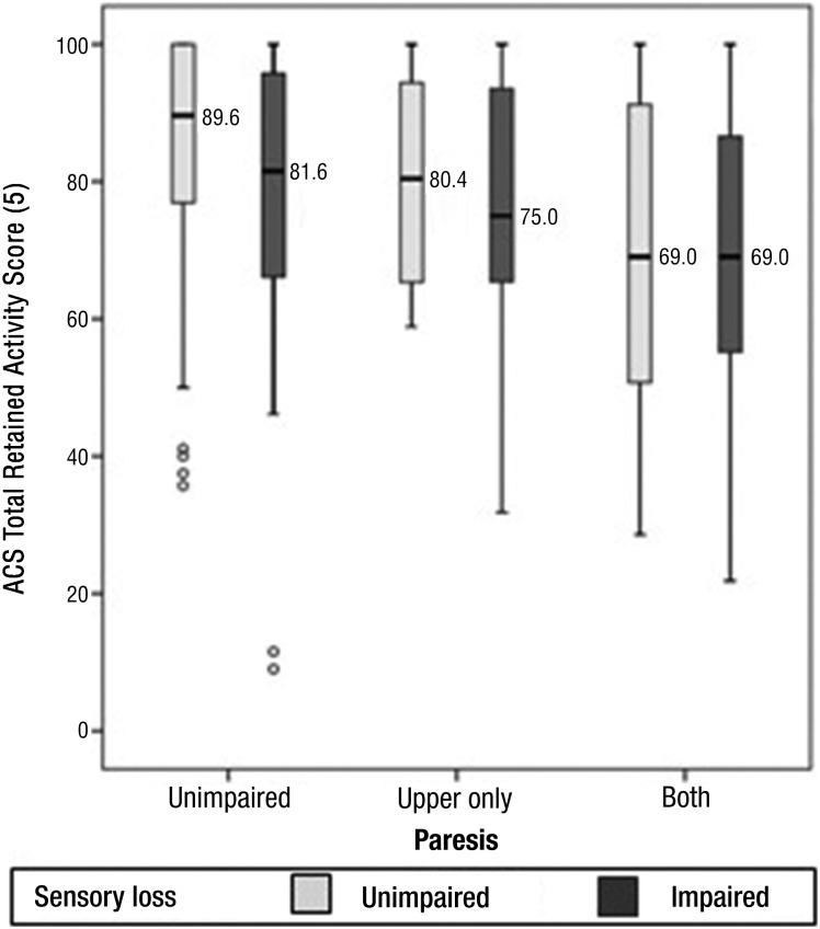
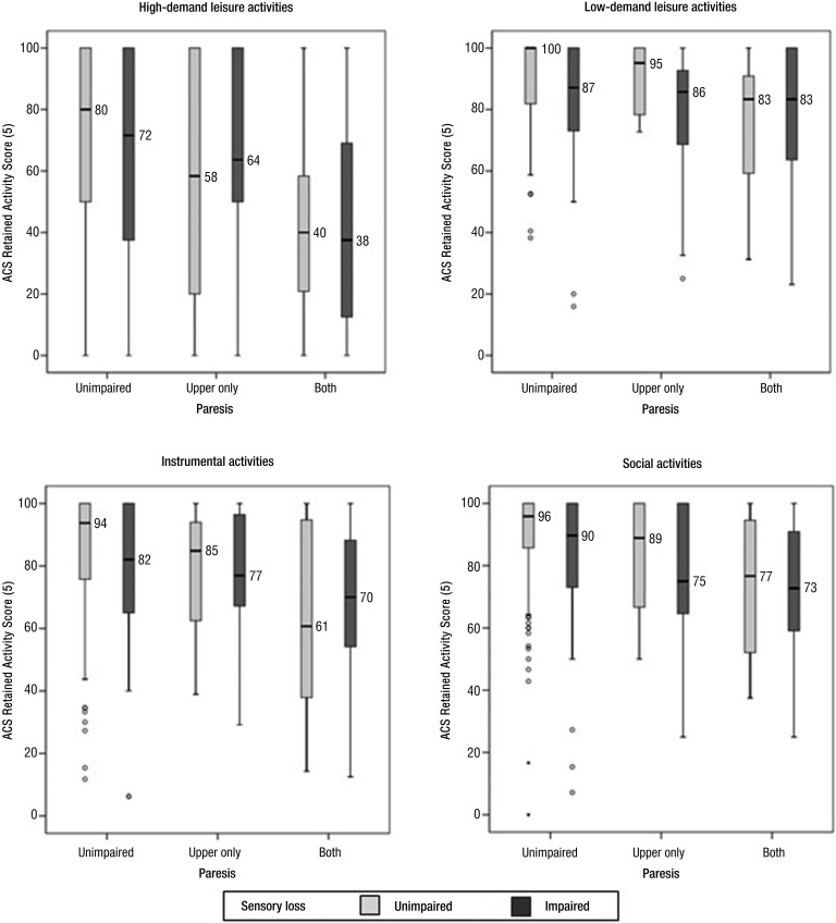
Method: Participants (N = 268) were assessed at hospital admission for somatosensory and motor impairment using the National Institutes of Health Stroke Scale. Participation was assessed using the Activity Card Sort (ACS) in the postacute phase. Between-group differences in activity participation were analyzed for participants with and without somatosensory impairment and with or without paresis.
Results: Somatosensory impairment was experienced in 33.6% of the sample and paresis in 42.9%. ACS profiles were obtained at a median of 222 days poststroke. Somatosensory loss alone (z = 1.96, p = .048) and paresis in upper and lower limbs without sensory loss (z = 4.62, p < .001) influenced activity participation.
Conclusion: Somatosensory impairment is associated with reduced activity participation; however, paresis of upper and lower limbs can mask the contribution of sensory loss.
Copyright © 2018 by the American Occupational Therapy Association, Inc.
Figures
References
-
- Baum C. M., & Edwards D. F. (2001). Activity Card Sort (ACS): Test manual. St. Louis: Program in Occupational Therapy, Washington University School of Medicine.
-
- Blennerhassett J. M., Carey L. M., & Matyas T. A. (2008). Clinical measures of handgrip limitation relate to impaired pinch grip force control after stroke. Journal of Hand Therapy, 21, 245–253. https://doi.org/10.1197/j.jht.2007.10.021 - DOI - PubMed
-
- Blennerhassett J. M., Matyas T. A., & Carey L. M. (2007). Impaired discrimination of surface friction contributes to pinch grip deficit after stroke. Neurorehabilitation and Neural Repair, 21, 263–272. https://doi.org/10.1177/1545968306295560 - DOI - PubMed
-
- Borstad A. L., & Nichols-Larsen D. S. (2014). Assessing and treating higher level somatosensory impairments post stroke. Topics in Stroke Rehabilitation, 21, 290–295. https://doi.org/10.1310/tsr2104-290 - DOI - PubMed
-
- Brott T., Adams H. P. Jr., Olinger C. P., Marler J. R., Barsan W. G., Biller J., . . . Hertzberg V. (1989). Measurements of acute cerebral infarction: A clinical examination scale. Stroke, 20, 864–870. https://doi.org/10.1161/01.STR.20.7.864 - DOI - PubMed
Publication types
MeSH terms
LinkOut - more resources
Full Text Sources
Other Literature Sources
Medical