. 2018 Jun:519:106-120.
doi: 10.1016/j.virol.2018.04.007. Epub 2018 Apr 22.

Macaque homologs of Kaposi's sarcoma-associated herpesvirus (KSHV) infect germinal center lymphoid cells, epithelial cells in skin and gastrointestinal tract and gonadal germ cells in naturally infected macaques

Affiliations

Macaque homologs of Kaposi's sarcoma-associated herpesvirus (KSHV) infect germinal center lymphoid cells, epithelial cells in skin and gastrointestinal tract and gonadal germ cells in naturally infected macaques

Helle Bielefeldt-Ohmann et al. Virology. 2018 Jun.

Abstract

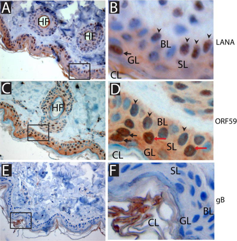
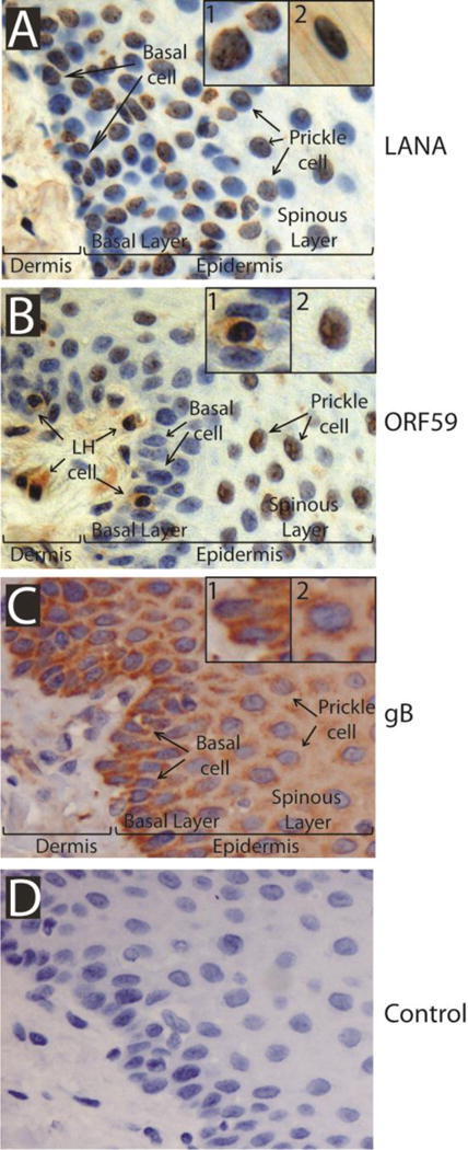
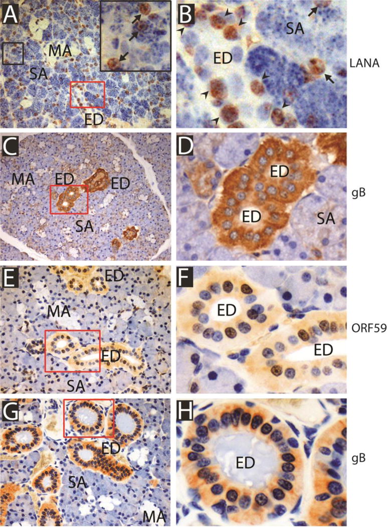
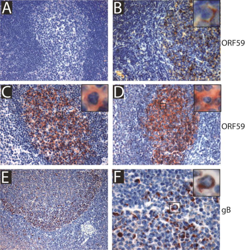
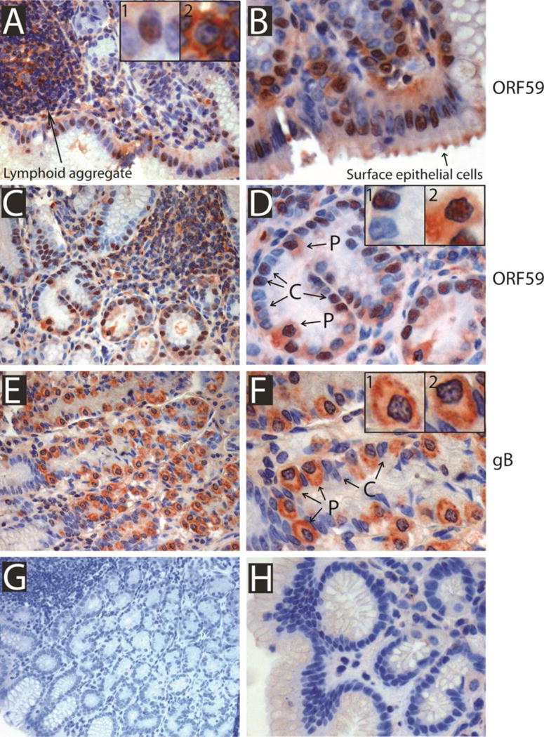
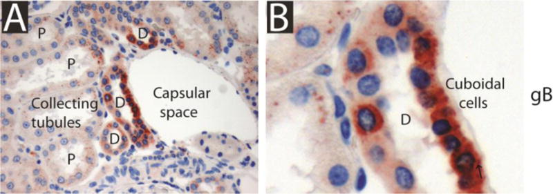
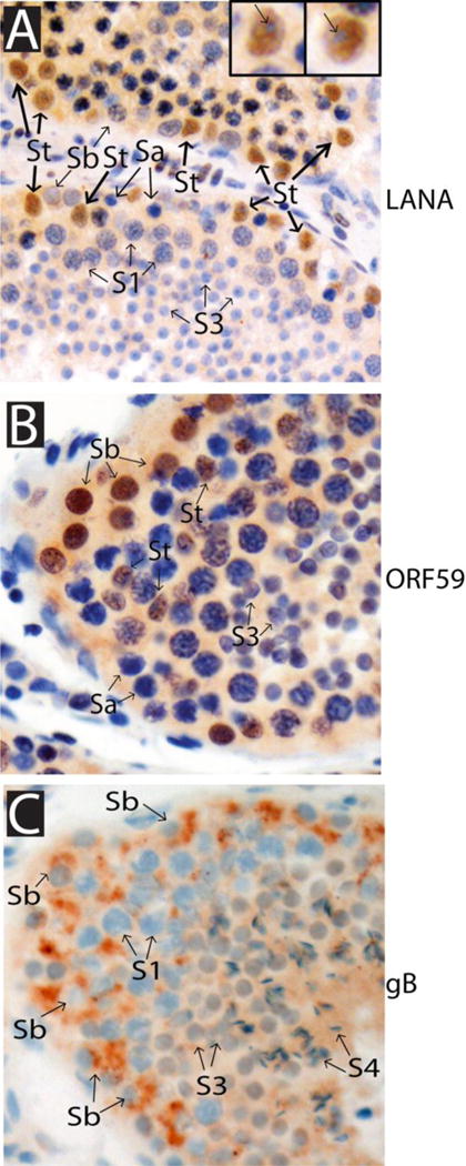
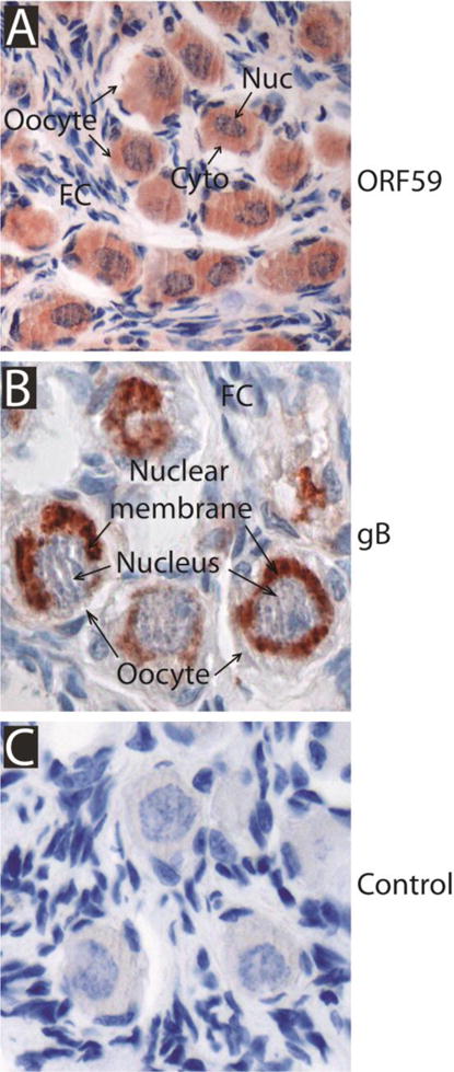
We developed a set of rabbit antisera to characterize infections by the macaque RV2 rhadinovirus homologs of KSHV. We analyzed tissues from rhesus and pig-tailed macaques naturally infected with rhesus rhadinovirus (RRV) or Macaca nemestrina rhadinovirus 2 (MneRV2). Our study demonstrates that RV2 rhadinoviruses have a tropism for epithelial cells, lymphocytes and gonadal germ cells in vivo. We observed latent infections in both undifferentiated and differentiated epithelial cells with expression of the latency marker, LANA. Expression of the early (ORF59) and late (glycoprotein B) lytic markers were detected in highly differentiated cells in epithelial ducts in oral, renal, dermal and gastric mucosal tissue as well as differentiated germ cells in male and female gonads. Our data provides evidence that epithelial and germ cell differentiation in vivo induces rhadinovirus reactivation and suggests that infected epithelial and germ cells play a role in transmission and dissemination of RV2 rhadinovirus infections in vivo.

Keywords: Epithelial; Germ cell; Immunohistochemistry; KSHV; Lymphocyte; Macaque; MneRV2; RRV; Rhadinovirus.


Figures

Fig. 1
Fig. 1. RV2 rhadinovirus infection in stratified skin epithelium
Formalin-fixed skin tissue from an adult SRV-2 positive pig-tailed macaque (F94292) from the WaNPRC was immuno-labeled with the 4109 rabbit panRV2 anti-LANA antiserum (A, B), the 425 rabbit panRV2 anti-ORF59 antiserum (C, D) or the 1508 rabbit panRV1/RV2 anti-gB antiserum (E, F) as described in Materials and Methods. Magnifications of the regions boxed in panels A, C and E are shown in panels B, D and F. HF = hair follicle; GL = granular layer; SL = spinous layer; BL = basal layer; CL = cornified layer. Arrowheads indicate columnar epithelial cells in the basal layer; red arrows indicate cells in the spinous layer with round nuclei; black arrows indicate cells in the granular layer with a squamous appearance.
Fig 2
Fig 2. RV2 rhadinovirus infection in stratified gingival mucosal epithelium
Formalin-fixed gingival tissue from an adult pig-tailed macaque (K01055) diagnosed with SIV-induced AIDS was immuno-labeled with 4109 panRV2 anti-LANA antiserum (A), 425 panRV2 anti-ORF59 antiserum (B), 1508 panRV1/RV2 anti-gB antiserum (C) or rabbit pre-immune serum (D). The position of the dermis/epidermis junction is indicated and the approximate positions of the basal and spinous layers of the stratified gingival epithelium are shown. Langerhans cells (LH) in the dermal layer, columnar basal cells in the basal layer and rounded prickle cells in the spinous layer are marked. A1 inset panel = rounded prickle cell with staining for RV2 LANA in the nucleus; A2 inset panel = flattened cell in the granular layer with nuclear RV2 LANA staining; B1 insert panel = Langerhans cell traversing the dermal/epidermal junction with ORF59 expression in both the nucleus and cytoplasm. Adjacent columnar basal cells are negative; B2 insert panel = prickle cell in the spinous layer with strong ORF59 expression in the nucleus but no cytoplasmic labeling; C1 insert panel = columnar basal cell with strong cytoplasmic and perinuclear gB expression; C2 insert panel = prickle cell with strong cytoplasmic and perinuclear gB expression.
Fig 3
Fig 3. RV2 rhadinovirus infection in salivary mucosal epithelium
Formalin-fixed salivary gland tissues from an adult pig-tailed macaque (K01055) diagnosed with SIV-induced AIDS (A-D) and an adult rhesus macaque (A01110) diagnosed with AIDS-associated lymphoma (E-H) were reacted with the 4109 panRV2 anti-LANA antiserum (A, B), the 425 anti-ORF59 antiserum (E, F) or the 1508 panRV1/RV2 anti-gB antiserum (C, D, G, H). Panel A: the small region boxed in black is magnified in the upper right box. The small regions boxed in red in panels A,C, E and G are magnified in panels B, D, F and H, respectively. Arrows in panels A and B point to LANA positive nuclei in serous acini (SA) and mucous acini (MA); arrowheads in panel B point to LANA positive nuclei in epithelial cells lining the excretory duct (ED).
Fig 4
Fig 4. RV2 rhadinovirus infection in lymphoid tissue
Formalin-fixed lymphoid tissue from a 6 month old juvenile pig-tailed macaque (M03105) (A), the 6 month old juvenile pig-tailed macaque (M03011) perinatally infected with SIV (B) and the adult SRV-2 positive pig-tailed macaque (F94292) (C-F) were immuno-labeled with the 425 panRV2 anti-ORF59 antiserum (A-D) or the 1508 panRV1/RV2 anti-gB antiserum (E, F). Lymph node (A-C); spleen (D-F). The single magnified cells show the cytoplasmic ORF59 staining (B-D) and cytoplasmic gB staining (F).
Fig 5
Fig 5. RV2 rhadinovirus infection in gastric mucosal epithelium
Formalin-fixed gastric tissue from a SHIV-positive six month old juvenile pig-tailed macaque that been part of a maternal SHIV transmission study (M03011; A, B), an adult pig-tailed macaque (K01055) diagnosed with SIV-induced AIDS (C-F) and a SHIV-negative six month old juvenile pig-tailed macaque (M03105; G-H) were immuno-labeled with the 425 panRV2 anti-ORF59 antiserum (A-D, H) or the 1508 panRV1/RV2 anti-gB antiserum (E-G). The positions of a lymphoid aggregate, surface epithelial cells and chief (C) and parietal (P) cells of the gastric pit are indicated. A1 inset panel shows the nuclear ORF59 expression in a surface epithelial cell; A2 inset panel shows the cytoplasmic ORF59 expression in lymphocytes within the lymphoid aggregate; D1 inset panel shows the nuclear ORF59 expression in the chief cells lining the gastric pit; D2 inset panel shows the nuclear and cytoplasmic ORF59 expression in the parietal cells lining the gastric pit; F1 and F2 inset panels show the cytoplasmic and perinuclear gB expression in the parietal cells. Panels B, D and F are magnified regions of the tissue sections shown in A, C and E, respectively.
Fig 6
Fig 6. RV2 rhadinovirus infection in cuboidal epithelial cells in the kidney
Formalin-fixed kidney tissue from an adult rhesus macaque (02181) with SHIV-induced AIDS-lymphoma was immuno-labeled with the 1508 panRV1/RV2 anti-gB antiserum. The positions of the capsular space and the distal (D) and proximal (P) collecting tubules are shown. The gB-positive cuboidal epithelial cells lining the distal tubule are magnified in B.
Fig 7
Fig 7. RV2 rhadinovirus infection in the testes
Formalin-fixed testicular tissues from adult male rhesus macaques A) A01112 and B,C) A01110 diagnosed with SIV-induced AIDS-lymphoma were immuno- labeled with the 4109 panRV2 anti-LANA antiserum (A), the 425 panRV2 anti-ORF59 antiserum (B) or the 1508 panRV1/RV2 anti-gB antiserum (C). The positions of Sertoli cells (St), undifferentiated Type A spermatogonial stem cells (Sa), immature Type B spermatogonia (Sb), primary spermatocyte (S1), spermatid (S3), spermatozoa (S4) are indicated. Panel A inserts: LANA-positive Sertoli cells showing absence of LANA-expression in nucleoli.
Fig 8
Fig 8. RV2 rhadinovirus infection in ovarian tissue
Formalin-fixed ovarian tissue from an adult female SRV-2-infected pig-tailed macaque (F94292) was immuno-labeled with either the 425 panRV2 anti- ORF59 antiserum (A), the 1508 panRV1/RV2 anti-gB antiserum (B), or pre-immune rabbit serum (C). The positions of the follicular cells (FC) and oocytes are shown with positions of the nucleus (Nuc), nuclear membrane and cytoplasm (Cyto).

Similar articles

Cited by

References

    1. Abe Y, Matsubara D, Gatanaga H, Oka S, Kimura S, Sasao Y, Saitoh K, Fujii T, Sato Y, Sata T, Katano H. Distinct expression of Kaposi’s sarcoma-associated herpesvirus-encoded proteins in Kaposi’s sarcoma and multicentric Castleman’s disease. Pathol Int. 2006;56:617–624. - PubMed
    1. Akula SM, Pramod NP, Wang FZ, Chandran B. Integrin alpha3beta1 (CD 49c/29) is a cellular receptor for Kaposi’s sarcoma-associated herpesvirus (KSHV/HHV-8) entry into the target cells. Cell. 2002;108:407–419. - PubMed
    1. Alexander L, Denekamp L, Knapp A, Auerbach MR, Damania B, Desrosiers RC. The primary sequence of rhesus monkey rhadinovirus isolate 26-95: sequence similarities to Kaposi’s sarcoma- associated herpesvirus and rhesus monkey rhadinovirus isolate 17577. J Virol. 2000;74:3388–3398. - PMC - PubMed
    1. Alvisi G, Marin O, Pari G, Mancini M, Avanzi S, Loregian A, Jans DA, Ripalti A. Multiple phosphorylation sites at the C-terminus regulate nuclear import of HCMV DNA polymerase processivity factor ppUL44. Virology. 2011;417:259–267. - PubMed
    1. Auerbach MR, Czajak SC, Johnson WE, Desrosiers RC, Alexander L. Species specificity of macaque rhadinovirus glycoprotein B sequences. J Virol. 2000;74:584–590. - PMC - PubMed

Publication types

MeSH terms

LinkOut - more resources