Review
. 2018 Apr 21;11(2):35.
doi: 10.3390/ph11020035.

Research Progress on Rolling Circle Amplification (RCA)-Based Biomedical Sensing

Affiliations
Review

Research Progress on Rolling Circle Amplification (RCA)-Based Biomedical Sensing

Lide Gu et al. Pharmaceuticals (Basel). .

Abstract

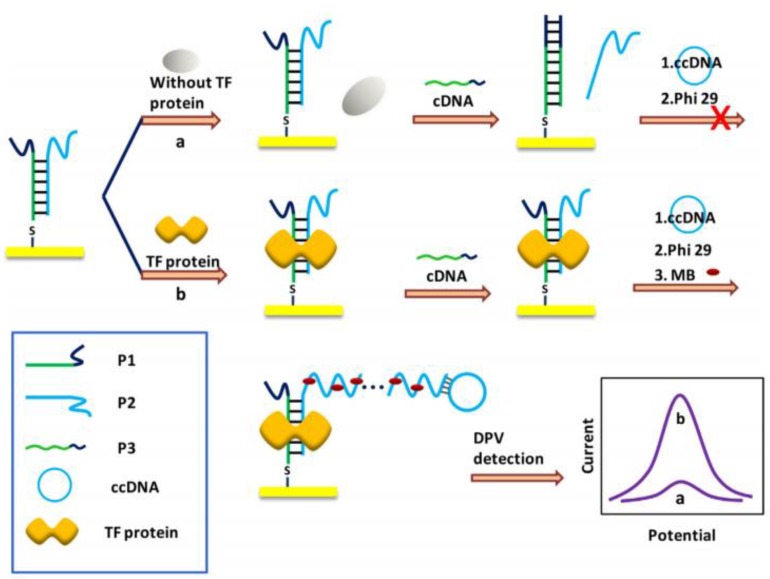
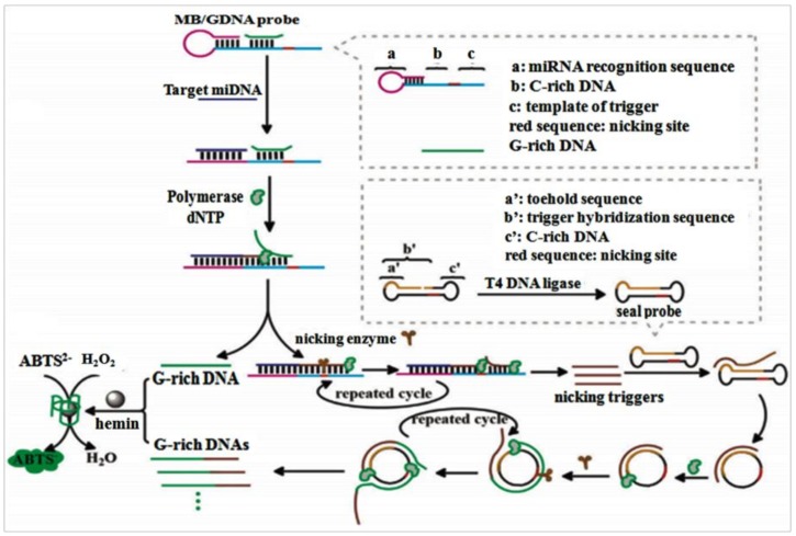
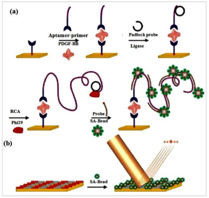
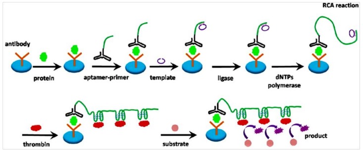
Enhancing the limit of detection (LOD) is significant for crucial diseases. Cancer development could take more than 10 years, from one mutant cell to a visible tumor. Early diagnosis facilitates more effective treatment and leads to higher survival rate for cancer patients. Rolling circle amplification (RCA) is a simple and efficient isothermal enzymatic process that utilizes nuclease to generate long single stranded DNA (ssDNA) or RNA. The functional nucleic acid unit (aptamer, DNAzyme) could be replicated hundreds of times in a short period, and a lower LOD could be achieved if those units are combined with an enzymatic reaction, Surface Plasmon Resonance, electrochemical, or fluorescence detection, and other different kinds of biosensor. Multifarious RCA-based platforms have been developed to detect a variety of targets including DNA, RNA, SNP, proteins, pathogens, cytokines, micromolecules, and diseased cells. In this review, improvements in using the RCA technique for medical biosensors and biomedical applications were summarized and future trends in related research fields described.

Keywords: biosensor; cancer; clinical diagnostics; rolling circle amplification (RCA).


Conflict of interest statement

The authors declare no conflict of interest.

Figures

Figure 1
Figure 1
Schematic diagram of G-quadruplex DNAzyme-based DNA ligase sensor. (a) The CT composed of three parts. Part I and III could hybridize with LT and form a split ring. Part II was a C-rich area where G-rich sequences are generated to form G-quadruplex structures after RCA. The split was repaired when T4 DNA ligase was introduced; then, LT was excised by Exo I and Exo III After annealing, PR was adhered to the circular template and activated RCA with Phi29 polymerase. Numerous G-rich sequences will be produced to fold into G-quadruplex units and bind hemin to form catalytic G-quadruplex DNAzymes, which can catalyze the oxidation of ABTS2− by H2O2 to ABTS, enhancing the absorption signal. (b) When the PNKP-triggered 5′-phosphroylation step was added to the substrate DNA, a PNKP sensor was easily designed based on the above sensing strategy. (LT, linear template; CT, circular template; ABTS, 2,2′-azino-bis(3-ethylbenzothiazoline-6-sulfonic acid)) [47].
Figure 2
Figure 2
Schematic diagram of AuNP-RCA sensor for multiple pathogens detection. (A) The PLP was designed as 5 regions which contain target-complementary sequences at the 5′ and 3′ ends (T1 and T2); the CPs hybridizes with a sequence-specific region (S); a Hpal restriction endonuclease digestion site (R) and the AuNP-MP binding sequences: a general region (G). (B) The scheme of detection process. (1) Linear PLP and target sequences were hybridized and linked to form circular template. (2) Immobilizing biotin-secondary antibody on the chip surface and incubated with streptavidin-Au nanoparticles and biotin-capture probe. (3) Circular PLP was added and hybridized with CPs to activate the RCA reaction, which can produce abundant binding sites for AuNP-MP and output signal of SPR [56].
Figure 3
Figure 3
Scheme of the aptasensor design. In the lack of target molecule, the ligation-RCA step was inhibited because complementary DNA (cDNA) was hybridized to the aptamer probe to generate a double-stranded duplex. Conversely, when the target molecule was introduced, the aptamer probe bound to the target molecule with high selectivity. As a result, the cDNA hybridized with the PLP insead of the aptamer probe. The PLP was circularized by DNA T4 ligase for RCA with Phi29 DNA polymerase. The product then hybridized with the loop of molecular beacons and generated a distinct fluorescence signal [64].
Figure 4
Figure 4
Schematic diagram of micromolecular biosensor based on electrochemistry coupled to RCA. (A) Formulation of RCA process based on structure-switching aptamer and sticky ends-based ligation. (a) Right part probe: target-aptamer binding, (b) left part probe: DNA denaturing to form a hairpin structure, (c) double probes were linked by E. coli DNA ligase with the same sticky ends, (d) the primer was adhered to the circular template by annealing, (e) RCA was initiated by adding Phi29 DNA polymerase and dNTPs. (B) Fabrication of the electrochemical biosensor. (a) Capture probes were anchored to the bare electrode, (b) blocking the electrode with MCH, (c) capture probe–RCA product hybridization for EIS quantitative determination [71].
Figure 5
Figure 5
Schematic diagram of TF sensing. Three ss-DNA probes were designed for the TF biosensor. P1 was attached to the electrode surface and hybridized with P2 to form a partially complementary dsDNA. The complementary part included a TF binding sites while the non-complementary part of P2 paired with RCA primer sequence. (a) In the absence of TF protein, P3 was displaced by P2 and hybridized with P1 to form a dsDNA strand, thus stopping RCA. (b) In the presence of TF protein, a stable TF-DNA complex formed and inhibited the displacement of P3, and then, initiated the RCA with ccDNA and Phi29 DNA polymerase. Finally, the electrochemical redox probe MB was combined with the RCA product and produced the signal that was detected by DPV [33].
Figure 6
Figure 6
Schematic diagram of the functional nucleic acid-based amplification system for miRNA detection. The MB/ GDNA probe were composed of four domains (see right frame). The hybridization was achieved between target miRNA and domain (a); then, the target miRNA was extended to domain (b) and domain (c) to form a complete duplex by Phi29 DNA polymerase (adhered to the domain b). Then, the duplex nicking site was recognized by the nicking enzyme specifically, and the extended DNA strand was cleaved at domain (c). The TIRCA was triggered when the seal probe was annealed with the primer which were nicking triggers and released by the circulation of the extension and cleavage processes at domain (c). When the toehold domain of seal probe was bound with trigger, the spontaneous branch migration leaded to an activated circular form, and the RCA was initiated. The resultant DNA duplex was cleaved at the nicking site in the circle by nicking enzyme. Hundreds of tandem repeats of GDNA was produced as the primers were extended by Phi29 DNA polymerase. Consequently, once the presence of hemin, the colorimetric detection was amplified by the formation of DNAzyme [99].
Figure 7
Figure 7
(a) Scheme of RCA-based microbead combines with aptamers for detection assay. Streptavidin coated periodic patterns links a biotinylated anti-PDGF-B specific aptamer. PDGF-BB was added and bound by the aptamer. An aptamer-primer complex recognized and bound to the protein. The primer tail of the aptamer was hybridized by a padlock probe, thereby initiating the RCA. The elongated concatemers hybridized with biotinylated probes which bound with streptavidin conjugated beads. (b) The diffraction gratings were formed by self-assembled streptavidin (SA)-coated beads on the RCA-based micropattern. The diffraction modes yielded when the illumination with a laser carried out [107].
Figure 8
Figure 8
Schematic diagram of using RCA coupled with thrombin catalysis to detect protein. Microplate coated with antibody which captured the target protein. Then, the aptamer-primer complex bound with the protein. The template hybridized with the primer and was circularized by ligase, and it encoded with a complementary sequence of the aptamer for thrombin. Subsequently, the RCA initiated and a long single-stranded DNA sequence was produced for thrombin. The generated ssDNA bound with thrombin molecules, achieving multiple thrombin labeling in sandwich complex. Thrombin catalyzes small peptide substrates into detectable product [110].
Figure 9
Figure 9
Scheme of RCA and electrochemical detection of DNA. (A) MBs connected with capture DNA1, and target DNA was added and tethered with it. Then, the primer DNA connected to the target DNA and the padlock probe after annealing. The RCA process was initiated to yield a long ssDNA after the padlock probe was circularized by ligase. Consequently, the long RCA products was digested to generate a crowd of short single ssDNA with same sequence as transfer DNA (t DNA) after Taq I DNA enzymes were added. (B) The surface of the Au electrode was covered by self-assembled DNA 2. And the electrochemical biosensor was formed by t DNA and capture DNA 2. The signal DNA loaded on Au NPs and hybridize with t DNA. The differential pulse voltammetry (DPV) scan monitored the quantity of the t DNA [116].
Figure 10
Figure 10
Scheme of the Ramos cells and aptamer reaction. Carboxyl-group-coated MBs connected with amino-modified aptamers of Ramos cells to form MB-DNA bio-complex which hybridized with cDNA. The bio-complex could instead of cDNA because of the stronger affinity between the aptamers and its targets when the presence of Ramos cells [116].
Figure 11
Figure 11
Schematic of a fluorescence biosensor for FR determination, based on HRCA and terminal protection. (A) FR connected with FA-ssDNA through FR-FA interaction and prevented the hydrolyzation of Exo I. Then, the compound hybridized with padlock probe, and formed a circular padlock probe with the aid of DNA ligase. Later, the HRCA reaction was activated by Bst DNA polymerase, P1, P2, and dNTPs to launch chain extensions and strand displacements, resulting a mass of long double-strand DNA (dsDNA) and ssDNA yielded. High fluorescence signal was monitored after introducing SYBR Green I. (B) In the absence of the target, ssDNA were hydrolyzed by Exo I and the HRCA was inhibited, so only weak fluorescence signal was detected [118].

Similar articles

Cited by

References

    1. Nilsson M., Gullberg M., Dahl F., Szuhai K., Raap A.K. Real-time monitoring of rolling-circle amplification using a modified molecular beacon design. Nucleic Acids Res. 2002;30:e66. doi: 10.1093/nar/gnf065. - DOI - PMC - PubMed
    1. Liu X., Xue Q., Ding Y., Zhu J., Wang L., Jiang W. Cascade Signal Amplification Strategy for Sensitive and Label-free DNA Detection Based on Exo III-catalyzed Recycling Coupled with Rolling Circle Amplification. Analyst. 2014;139:2884–2889. doi: 10.1039/C4AN00389F. - DOI - PubMed
    1. Li Y., Zeng Y., Ji X., Li X., Ren R. Cascade signal amplification for sensitive detection of cancer cell based on self-assembly of DNA scaffold and rolling circle amplification. Sens. Actuators B Chem. 2012;171–172:361–366. doi: 10.1016/j.snb.2012.04.060. - DOI
    1. Khan S.A. Plasmid rolling-circle replication: Highlights of two decades of research. Plasmid. 2005;53:126–136. doi: 10.1016/j.plasmid.2004.12.008. - DOI - PubMed
    1. Zhao W., Cui C.H., Bose S., Guo D., Shen C., Wong W.P., Halvorsen K., Farokhzad O.C., Teo G.S.L., Phillips J.A., et al. Bioinspired multivalent DNA network for capture and release of cells. Proc. Natl. Acad. Sci. USA. 2012;109:19626–19631. doi: 10.1073/pnas.1211234109. - DOI - PMC - PubMed