Review
. 2018 Apr 23;8(2):20.
doi: 10.3390/biom8020020.

Protein⁻Phospholipid Interaction Motifs: A Focus on Phosphatidic Acid

Affiliations
Review

Protein⁻Phospholipid Interaction Motifs: A Focus on Phosphatidic Acid

Emeline Tanguy et al. Biomolecules. .

Abstract

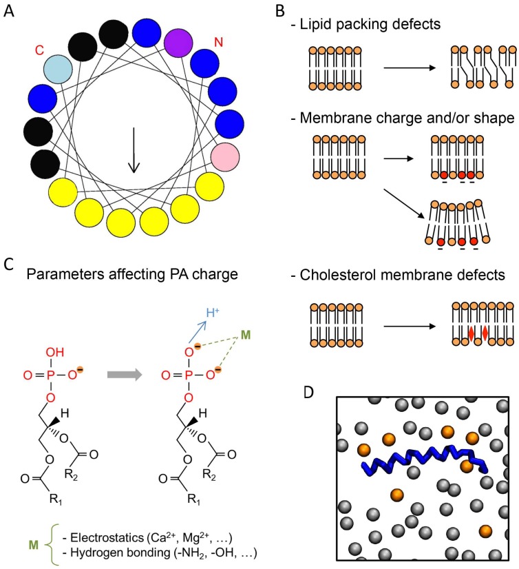
Cellular membranes are composed of thousands of different lipids usually maintained within a narrow range of concentrations. In addition to their well-known structural and metabolic roles, signaling functions for many lipids have also emerged over the last two decades. The latter largely depend on the ability of particular classes of lipids to interact specifically with a great variety of proteins and to regulate their localization and activity. Among these lipids, phosphatidic acid (PA) plays a unique role in a large repertoire of cellular activities, most likely in relation to its unique biophysical properties. However, until recently, only incomplete information was available to model the interaction between PA and its protein partners. The development of new liposome-based assays as well as molecular dynamic simulation are now providing novel information. We will review the different factors that have shown to modulate the capacity of PA to interact with specific domains in target proteins.

Keywords: interaction motif; lipid binding; membrane; phosphatidic acid; phospholipase D.


Conflict of interest statement

The authors declare no conflict of interest.

Figures

Figure 1
Figure 1
Typical phosphatidic acid (PA)-binding domain (PABD) and parameters affecting protein–PA interactions. (A) Amphipathic α-helix projection of a minimal characteristic PABD obtained with Heliquest software (http://heliquest.ipmc.cnrs.fr). Arrow indicates hydrophobic moment. Critical basic and hydrophobic residues are indicated in blue and yellow, respectively. Histidine (light blue), tryptophan (pink), and serine (purple) residues are also often present in the PABD. The amino-terminal (N) and carboxyl-terminal (C) regions of the PABD are indicated. (B) Different parameters affecting protein–PA binding are indicated on the right, including, from top to bottom: lipid-packing defects caused by fatty acyl unsaturation, negatively charged phospholipids such as phosphatidylserine (PS) (red) and membrane curvature, and cholesterol membrane defects (red diamond). Note that any of these parameters can affect PA binding either individually or in combination. Although cholesterol affects the hydrophobic core of the membrane or its hydrophilic interface, it is likely that the most important effect of cholesterol regarding the interaction of proteins with PA involves protein accessibility to hydrophilic headgroups of the membrane. (C) The net charge of PA is also modulated by different parameters such as ionic concentration and pH (adapted from [15]). The fatty acyl chain length and degree of unsaturation in the sn-1 or sn-2 positions (R1 and R2) also modify the binding to PABDs. (D) Top view of the final shot of a molecular dynamics simulation (500 ns) of the PABD of Spo20p (blue) in a phosphatidylcholine (PC)/PA (grey/orange) artificial membrane [16]. Only the phosphate headgroups of the lipids of a single bilayer are shown.

References

    1. Arora A., Tamm L.K. Biophysical approaches to membrane protein structure determination. Curr. Opin. Struct. Biol. 2001;11:540–547. doi: 10.1016/S0959-440X(00)00246-3. - DOI - PubMed
    1. Basso L.G.M., Mendes L.F.S., Costa-Filho A.J. The two sides of a lipid-protein story. Biophys. Rev. 2016;8:179–191. doi: 10.1007/s12551-016-0199-5. - DOI - PMC - PubMed
    1. Shenoy S., Shekhar P., Heinrich F., Daou M., Gericke A., Ross A.H., Lösche M. Membrane association of the PTEN tumor suppressor: Molecular details of the protein–membrane complex from SPR binding studies and neutron reflection. PLoS ONE. 2012;7:e32591. doi: 10.1371/journal.pone.0032591. - DOI - PMC - PubMed
    1. Raja M., Spelbrink R.E.J., de Kruijff B., Killian J.A. Phosphatidic acid plays a special role in stabilizing and folding of the tetrameric potassium channel KcsA. FEBS Lett. 2007;581:5715–5722. doi: 10.1016/j.febslet.2007.11.039. - DOI - PubMed
    1. Ammar M.R., Kassas N., Bader M.F., Vitale N. Phosphatidic acid in neuronal development: A node for membrane and cytoskeleton rearrangements. Biochimie. 2014;107:51–57. doi: 10.1016/j.biochi.2014.07.026. - DOI - PubMed

Publication types

LinkOut - more resources