. 2018 Apr 23;7(4):32.
doi: 10.3390/cells7040032.

CDK5RAP2 Is an Essential Scaffolding Protein of the Corona of the Dictyostelium Centrosome

Affiliations

CDK5RAP2 Is an Essential Scaffolding Protein of the Corona of the Dictyostelium Centrosome

Valentin Pitzen et al. Cells. .

Abstract

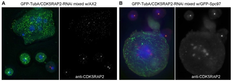
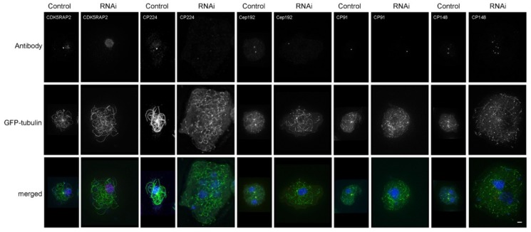
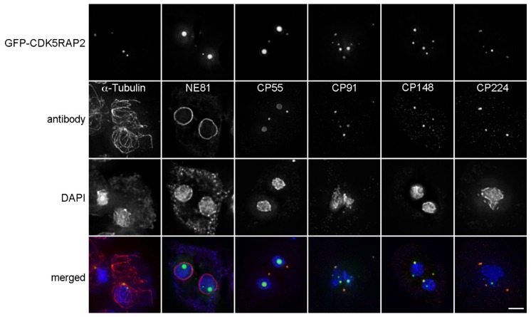
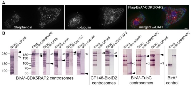
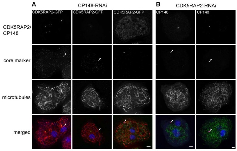
Dictyostelium centrosomes consist of a nucleus-associated cylindrical, three-layered core structure surrounded by a corona consisting of microtubule-nucleation complexes embedded in a scaffold of large coiled-coil proteins. One of them is the conserved CDK5RAP2 protein. Here we focus on the role of Dictyostelium CDK5RAP2 for maintenance of centrosome integrity, its interaction partners and its dynamic behavior during interphase and mitosis. GFP-CDK5RAP2 is present at the centrosome during the entire cell cycle except from a short period during prophase, correlating with the normal dissociation of the corona at this stage. RNAi depletion of CDK5RAP2 results in complete disorganization of centrosomes and microtubules suggesting that CDK5RAP2 is required for organization of the corona and its association to the core structure. This is in line with the observation that overexpressed GFP-CDK5RAP2 elicited supernumerary cytosolic MTOCs. The phenotype of CDK5RAP2 depletion was very reminiscent of that observed upon depletion of CP148, another scaffolding protein of the corona. BioID interaction assays revealed an interaction of CDK5RAP2 not only with the corona markers CP148, γ-tubulin, and CP248, but also with the core components Cep192, CP75, and CP91. Furthermore, protein localization studies in both depletion strains revealed that CP148 and CDK5RAP2 cooperate in corona organization.

Keywords: Dictyostelium; centriole; centrosome; microtubules; mitosis.


Conflict of interest statement

The authors declare no conflict of interest. The founding sponsors had no role in the design of the study; in the collection, analyses, or interpretation of data; in the writing of the manuscript, or in the decision to publish the results.

Figures

Figure 1
Figure 1
Subcentrosomal distribution of CDK5RAP2. Immunofluorescence deconvolution microscopy of AX2 control cells with rabbit anti-CDK5RAP2, rat anti-CP55 and mouse anti-CP224. Secondary antibodies against rabbit, rat and mouse antibodies were conjugated with AlexaFluor 568, AlexaFluor 488, and Cy5, respectively and cells were fixed with methanol. (A) The same representative centrosome is shown in all three channels as indicated. (B) Shows the corresponding intensity distributions for CDK5RAP2 (green), CP55 (blue) and CP224 (red) along the line shown in the upper left of (A).
Figure 2
Figure 2
CDK5RAP2 is present at mitotic spindle poles. (A) Immunofluorescence microscopy of AX2 control cells in interphase and mitosis (as indicated) stained with rabbit anti-CDK5RAP2 and rat anti-α-tubulin. DNA staining with DAPI is shown in the merged image. Secondary antibodies were anti-rabbit AlexaFluor 488 and anti-rat AlexaFluor 568. Cells were fixed with glutaraldehyde. Maximum intensity projections of deconvolved images. To allow comparison of labeling intensities in mitotic vs. interphase cells, the maximum display intensity of the green channel was set using neighboring interphase cells. Bar = 2 µm. (B) Immunoblot of a nuclear extract of untransformed AX2 cells stained with anti-CDK5RAP2/anti-rabbit-alkaline phosphatase. Color detection was performed with nitroblue tetrazolium chloride (NBT) and bromo-chloro-indolyl-phosphate (BCIP).
Figure 3
Figure 3
GFP-CDK5RAP2 briefly disappears from the centrosome during prophase. Confocal spinning disk live cell imaging of a mitotic GFP-CDK5RAP2 cell. Selected time points from Video S1 are displayed. Mitotic centrosomes are highlighted by arrowheads. Cells were viewed under agar overlay [35]. Bar = 2 µm.
Figure 4
Figure 4
GFP-CDK5RAP2ki shows hardly any recovery after photobleaching. (A) Confocal spinning disk live cell imaging of GFP-CDK5RAP2ki cells. Selected time points from Video S2 are displayed. A maximum intensity projection of three confocal slices (z-distance 0.25 µm) is shown. The bleached centrosome is highlighted by an arrowhead. Cells were viewed under agar overlay [35]. Bar = 2 µm. (B) FRAP curve mean intensity values and error bars indicating SD (n = 10) are plotted against time.
Figure 5
Figure 5
CDK5RAP2RNAi cells contain display disrupted microtubule arrays and enlarged nuclei. Immunofluorescence microscopy of GFP-TubA/CDK5RAP2RNAi cells mixed with AX2 control cells (A) or GFP-Spc97 cells (B) stained with rabbit anti-CDK5RAP2. DNA-staining with DAPI is shown in the merged image. The secondary antibody was anti-rabbit-AlexaFluor568. Cells were fixed with glutaraldehyde. Maximum intensity projections of deconvolved images are displayed. Bar = 2 µm.
Figure 6
Figure 6
CDK5RAP2RNAi causes centrosome disruption. Immunofluorescence microscopy of GFP-TubA/CDK5RAP2RNAi cells stained with the indicated antibodies, in comparison to GFP-α-tubulin control cells. DNA staining with DAPI is shown in the merged images. Secondary anti-rabbit/rat/mouse antibodies were all AlexaFluor568. Cells were fixed with methanol. Maximum intensity projections of deconvolved images are displayed. Bar = 2 µm.
Figure 7
Figure 7
Overexpressed GFP-CDK5RAP2 is concentrated in discrete foci in the cytosol or nucleus. Immunofluorescence microscopy of GFP-CDK5RAP2 cells stained with the indicated antibodies. DNA was stained with DAPI. Secondary anti-rabbit/rat/mouse antibodies were conjugated with AlexaFluor 568. Cells were fixed with methanol. Maximum intensity projections of deconvolved images are displayed, except for the GFP-CDK5RAP2 sample co-stained for NE81 (lamin, nuclear envelope marker, [34]), where only the optical section through the center of the nucleus is shown, to show that there are indeed GFP-CDK5RAP2 foci inside the nucleus. Bar = 2 µm.
Figure 8
Figure 8
Centrosomal interactions of biotinylase-tagged CDK5RAP2 in the BioID assay. (A) Immunofluorescence microscopy ofFlag-BirA-tagged CDK5RAP2RNAi cells stained with anti-α-tubulin and streptavidin-AlexaFluor 488. DNA staining with DAPI is shown in the merged image. The secondary antibody was anti-rat AlexaFluor 568. Cells were fixed with glutaraldehyde. Maximum intensity projections of deconvolved images are displayed. Bar = 2 µm. (B) BioID Western blot analysis according to [30]. Centrosome extracts prepared from the cell lines indicated at the bottom were electrophoresed and blotted. Single lanes were cut from broad blots, stained individually with streptavidin-alkaline phosphatase (strep) or antibodies as indicated on top, and re-aligned after staining as shown. Bands were visualized using conjugates with alkaline phosphatase and NBT/BCIP color detection. Positions of the respective fusion proteins always showing self-biotinylation are marked by open arrowheads. Positions of endogenous, biotinylated proteins are labeled by filled arrowheads. The position of the double band representing biotinylated mitochondrial proteins is highlighted by an asterisk.
Figure 9
Figure 9
CDK5RAP2RNAi and CP148RNAi cause similar centrosome disruptions. Immunofluorescence microscopy of CDK5RAP2-GFP/CP148RNAi (A) and GFP-TubA/CDK5RAP2RNAi (B) cells stained with the indicated antibodies. DNA-staining with DAPI is shown in the merged images. In (A) primary and secondary antibodies were anti-α-tubulin/anti-rat-Atto647n and anti-Cep192 (core marker; arrowhead)/anti-rabbit-AlexaFluor 568, in (B) anti-CP55 (core marker; arrowhead)/anti-rat-AlexaFluor 568 and anti-CP148/anti-rabbit-Alexa 647. Cells were fixed with methanol. Maximum intensity projections of deconvolved images are displayed. Bars = 2 µm.

Similar articles

Cited by

References

    1. Fu J., Glover D. How the newborn centriole becomes a mother. Cell Cycle. 2016;15:1521–1522. doi: 10.1080/15384101.2016.1164566. - DOI - PMC - PubMed
    1. Novak Z.A., Wainman A., Gartenmann L., Raff J.W. Cdk1 phosphorylates Drosophila Sas-4 to recruit polo to daughter centrioles and convert them to centrosomes. Dev. Cell. 2016;37:545–557. doi: 10.1016/j.devcel.2016.05.022. - DOI - PMC - PubMed
    1. Fry A.M., Sampson J., Shak C., Shackleton S. Recent advances in pericentriolar material organization: Ordered layers and scaffolding gels. F1000Research. 2017;6:1622. doi: 10.12688/f1000research.11652.1. - DOI - PMC - PubMed
    1. Pedersen L.B., Schroder J.M., Satir P., Christensen S.T. The ciliary cytoskeleton. Compr. Physiol. 2012;2:779–803. - PubMed
    1. Devos D.P., Gräf R., Field M.C. Evolution of the nucleus. Curr. Opin. Cell Biol. 2014;28C:8–15. doi: 10.1016/j.ceb.2014.01.004. - DOI - PMC - PubMed

LinkOut - more resources