Review
. 2018 Apr 23;8(4):268.
doi: 10.3390/nano8040268.

ZnO Nanostructures for Drug Delivery and Theranostic Applications

Affiliations
Review

ZnO Nanostructures for Drug Delivery and Theranostic Applications

Marina Martínez-Carmona et al. Nanomaterials (Basel). .

Abstract

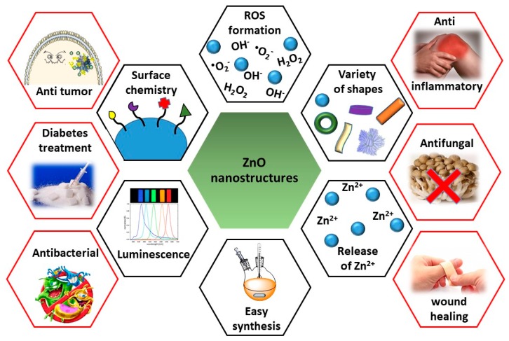
In the last two decades, zinc oxide (ZnO) semiconductor Quantum dots (QDs) have been shown to have fantastic luminescent properties, which together with their low-cost, low-toxicity and biocompatibility have turned these nanomaterials into one of the main candidates for bio-imaging. The discovery of other desirable traits such as their ability to produce destructive reactive oxygen species (ROS), high catalytic efficiency, strong adsorption capability and high isoelectric point, also make them promising nanomaterials for therapeutic and diagnostic functions. Herein, we review the recent progress on the use of ZnO based nanoplatforms in drug delivery and theranostic in several diseases such as bacterial infection and cancer.

Keywords: Quantum dots; ZnO nanoparticles; anti-inflammation; anti-tumour; antibacterial; antifungal; diabetes treatment; drug delivery; theranostic; wound healing.


Conflict of interest statement

The authors declare no conflict of interest.

Figures

Figure 1
Figure 1
Diagram summarizing the main characteristics of ZnO nanostructures (black hexagons) and their principal applications in biomedicine (red hexagons).
Figure 2
Figure 2
(a) Scheme of the multiple proposed effects of ZnO QDs as a multi-functional antitumour treatment. Reproduced with permission from [44]. American Chemical Society, 2017; (b) Scheme of the combined mechanism of action of DOX-FA-ZnO NS for breast carcinoma therapy. Reproduced with permission from [48]. Elsevier, 2017.
Figure 3
Figure 3
(a) Images of BxPC-3 tumour-bearing nude mice after 18 and 36 days under different treatments; (b) H&E staining of tumour slices after 36 days of treatments by different agents. Reproduced with permission from [34]. American Chemical Society, 2016.
Figure 4
Figure 4
(a) Synthesis scheme and mechanism of action of Fe3O4@ZnO:Er3+,Yb3+@β-CD nano-composites; (b) Graph of VP-16 release from the Fe3O4@ZnO:Er3+,Yb3+@(β-CD)–(VP-16) depending on the number of microwave cycles applied. Reproduced with permission from [55]. Elsevier, 2015.
Figure 5
Figure 5
(a) DOX release profiles from ZnO@MSNsDOX at 3 different pH values (7.4, 5.0, and 2.0); (b) In vitro viability of HeLa cells in the presence of COOHMSNs, ZnO@MSNs, ZnO@MSNsDOX, and free DOX. Reproduced with permission from [56]. American Chemical Society, 2011; (c) Confocal microscopy images taken at different times of A549 cells incubated with the MIT-loaded, ZnO-gated MCNs. Cell membranes were stained in green, cell nuclei were stained in blue and released drugs wre presented red. Reproduced with permission from [60]. Elsevier, 2016.
Figure 6
Figure 6
(a) Schematic representation of fabrication and degradation process of the ZnO@Dextran microgels; (b,c) Two different magnification SEM photographs of ZnO@Dextran microgels; (d) Digital photos of ZnO@Dextran microgels after incubation at pH 3.0 (left), 5.0 (middle) and 7.4 (right); (e) Cell viabilities of HeLa cells after being incubated with different samples under different conditions. Reproduced with permission from [61]. John Wiley and Sons, 2018.
Figure 7
Figure 7
Reduction in colony counts (from 5 × 10⁷) observed after exposure to increasing concentrations of: (a) H2O2; (b) ZnO-NSPs; or (c) ZnO-NPYs, with and without 50 mM NAC. Reproduced with permission from [83]. Royal Society of Chemistry, 2018.
Figure 8
Figure 8
Fluorescence micrographs of E. coli biofilms grown up onto ZnO NRs surfaces: (a) without; or (b) with light treatment. Alive cells were stained in green and dead cells in red; (c) Light-dose dependence cytotoxic effect on E. faecalis MSCL 302, L. monocytogenes ATCL3C 7644 and E. coli O157:H7 biofilms grown up onto ZnO NRs coated surfaces. Non-illuminated biofilms grown on plastic surfaces were used as control and effect the NRs in absence of light was expressed as dark toxicity. Reproduced with permission from [86].
Figure 9
Figure 9
(a) Image of isolated mouse intestinal loop 6 h after the injection of 1 µg of CT and 1.25 µg µL−1 of ZnO QDs or only CT in the control one; (b) Fluid accumulation (FA) ratio after 6 h of injection; (n = 10 mice with 15–20 loops studied per group). Reproduced with permission from [90]. Elsevier, 2016.
Figure 10
Figure 10
SEM images of (ad) Penicillium expansum and (eh) Botrytis cinerea without (a,b,e,f) or with (c,d,g,h) the treatment of ZnO QDs suspension. Reproduced with permission from [97]. Elsevier, 2011.
Figure 11
Figure 11
(a) Determination of iNOS and COX-2 mRNA levels by RT-PCR; (b) Determination of iNOS and COX-2 protein levels by Western blot (mean ± SD of n = 3 and * p < 0.05 versus LPS alone). Reproduced with permission from [119]. Elsevier, 2015.
Figure 12
Figure 12
In vivo images of the wound healing process in Sprague−Dawley rats. Reproduced with permission from [125]. American Chemical Society, 2012.

References

    1. Jia Z., Misra R.D.K. Tunable ZnO quantum dots for bioimaging: synthesis and photoluminescence. Mater. Technol. 2013;28:221–227. doi: 10.1179/1753555713Y.0000000061. - DOI
    1. Resch-Genger U., Grabolle M., Cavaliere-Jaricot S., Nitschke R., Nann T. Quantum dots versus organic dyes as fluorescent labels. Nat. Methods. 2008;5:763–775. doi: 10.1038/nmeth.1248. - DOI - PubMed
    1. Volokitin Y., Sinzig J., de Jongh L.J., Schmid G., Vargaftik M.N., Moiseevi I.I. Quantum-size effects in the thermodynamic properties of metallic nanoparticles. Nature. 1996;384:621–623. doi: 10.1038/384621a0. - DOI
    1. Asok A., Gandhi M.N., Kulkarni A.R. Enhanced visible photoluminescence in ZnO quantum dots by promotion of oxygen vacancy formation. Nanoscale. 2012;4:4943. doi: 10.1039/c2nr31044a. - DOI - PubMed
    1. Zhang Z.-Y., Xiong H.-M. Photoluminescent ZnO Nanoparticles and Their Biological Applications. Materials. 2015;8:3101–3127. doi: 10.3390/ma8063101. - DOI

LinkOut - more resources