Review
. 2018 Apr 23;19(4):1264.
doi: 10.3390/ijms19041264.

Molecular Pharmacology of VEGF-A Isoforms: Binding and Signalling at VEGFR2

Affiliations
Review

Molecular Pharmacology of VEGF-A Isoforms: Binding and Signalling at VEGFR2

Chloe J Peach et al. Int J Mol Sci. .

Abstract

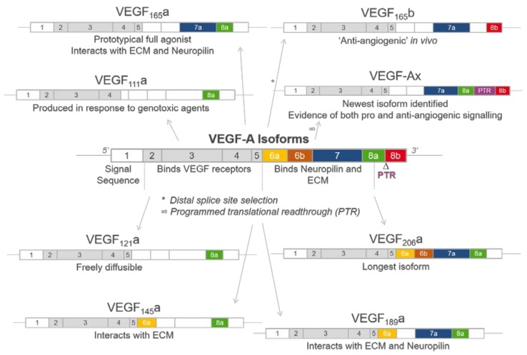
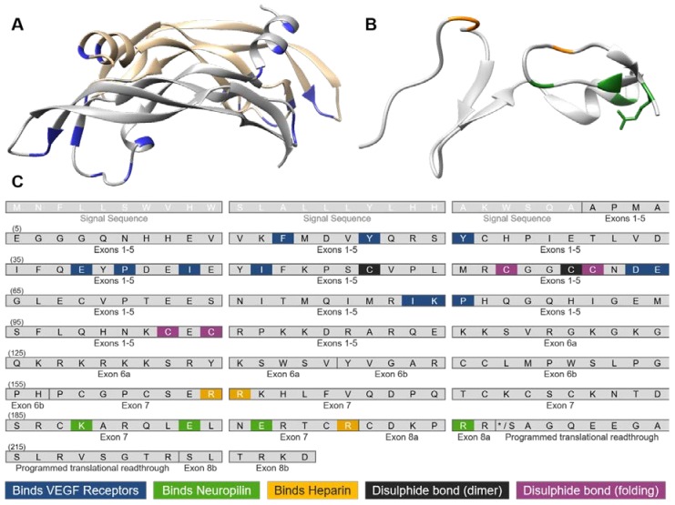
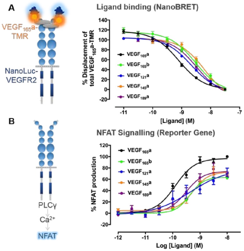
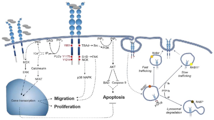
Vascular endothelial growth factor-A (VEGF-A) is a key mediator of angiogenesis, signalling via the class IV tyrosine kinase receptor family of VEGF Receptors (VEGFRs). Although VEGF-A ligands bind to both VEGFR1 and VEGFR2, they primarily signal via VEGFR2 leading to endothelial cell proliferation, survival, migration and vascular permeability. Distinct VEGF-A isoforms result from alternative splicing of the Vegfa gene at exon 8, resulting in VEGFxxxa or VEGFxxxb isoforms. Alternative splicing events at exons 5⁻7, in addition to recently identified posttranslational read-through events, produce VEGF-A isoforms that differ in their bioavailability and interaction with the co-receptor Neuropilin-1. This review explores the molecular pharmacology of VEGF-A isoforms at VEGFR2 in respect to ligand binding and downstream signalling. To understand how VEGF-A isoforms have distinct signalling despite similar affinities for VEGFR2, this review re-evaluates the typical classification of these isoforms relative to the prototypical, “pro-angiogenic” VEGF165a. We also examine the molecular mechanisms underpinning the regulation of VEGF-A isoform signalling and the importance of interactions with other membrane and extracellular matrix proteins. As approved therapeutics targeting the VEGF-A/VEGFR signalling axis largely lack long-term efficacy, understanding these isoform-specific mechanisms could aid future drug discovery efforts targeting VEGF receptor pharmacology.

Keywords: angiogenesis; blood vessel; endothelial cells; receptor tyrosine kinase inhibitors; splicing.


Conflict of interest statement

The authors declare no conflict of interest.

Figures

Figure 1
Figure 1
Schematic illustrating the structure of vascular endothelial growth factor A (VEGF-A) isoforms. The VEGF-A gene consists of eight exons, which can be alternatively spliced to generate a range of VEGF-A isoforms. These isoforms differ in length and have been designated VEGFxxx, where xxx represents the number of amino acids present. Each exon contains residues identified as conferring distinct properties if included in the resultant isoform, including VEGFR2, extracellular matrix (ECM) and Neuropilin (NRP) binding. A major site of alternative splicing occurs at exon 8, whereby proximal splicing results in the prototypical VEGFxxxa forms and distal splicing the “anti-angiogenic” VEGFxxxb isoforms containing exon 8b. Additionally, post translational read-through (PTR) using a non-canonical stop codon results in the VEGF-Ax isoform which contains a 22 amino acid extension in its C terminal domain.
Figure 2
Figure 2
Molecular structure of VEGF-A. (A) Anti-parallel homodimeric structure of VEGF-A encoded by exons 2–5 (PDB:1VPF), showing distinct VEGF monomers in grey and gold and residues interacting with VEGF receptors shown in blue; (B) C-terminus of VEGF165a is encoded by exons 7–8a (PDB:4DEQ), with residues that bind heparin (yellow) and Neuropilin-1 (green) highlighted; (C) Amino acid residues present in exons of the human VEGF-A sequence that interact with known binding partners. The open reading frame was derived from transcript NM_001025366.2 with exons denoted according to UniProt (P15692) and residues numbered according to residues in the final VEGF-A peptide following cleavage of the signal sequence. Based on published X-ray crystal structures, residues are highlighted that form non-covalent interactions with VEGFR1 [88], VEGFR2 [84], Neuropilin-1 [89] or heparin [70]. Cysteine residues forming intermolecular or intramolecular disulphide bonds, important for dimeric or folding structure, respectively, are also highlighted [26].
Figure 3
Figure 3
Quantifying VEGF-A isoform binding and downstream nuclear factor of activated T-cells (NFAT) signalling to derive pharmacological parameters. (A) Ligand binding affinities to VEGFR2 were quantified using HEK293 cells stably transfected with the full-length human VEGFR2 tagged at its N-terminus with the novel luciferase NanoLuc. Bioluminescence resonance energy transfer (BRET) experiments were then performed, whereby the close proximity of the donor NanoLuc tag with bound VEGF165a fluorescently with tetramethylrhodamine (VEGF165a-TMR) facilitates the non-radiative transfer of this energy to excite the acceptor TMR fluorophore which itself emits light at a longer wavelength. Cells were co-stimulated using a fixed concentration (3 nM) of single-site fluorescently labelled VEGF165a (VEGF165a-TMR) and increasing concentrations of competing unlabelled VEGF-A isoforms (60 min at 37 °C). These data were normalised to percentage displacement of VEGF165a-TMR alone and binding affinities (pKi values) of unlabelled isoforms estimated using the Cheng–Prusoff equation with VEGF165a-TMR Kd values calculated from previous saturation experiments (see [97] for more details). (B) Functional potencies of VEGF-A isoforms were derived from an NFAT reporter gene assay, whereby a Firefly luciferase inserted downstream of the NFAT promoter sequence was used to investigate the potency of unlabelled VEGF-A isoforms in respect to stimulating downstream NFAT production. HEK293 cells stably expressing full-length human VEGFR2 were stimulated with a concentration response course of unlabelled VEGF-A isoforms (5 h at 37 °C/5% CO2). A luminescence readout was indicative of NFAT production. All responses were expressed as a percentage of 10 nM VEGF165a. The potency and efficacy of VEGF-A isoforms in respect to NFAT production were calculated using non-linear least square regression. All data were pooled from 4/5 independent experiments and expressed as ± S.E.M. Figures modified from Kilpatrick et al. (2017) [97].
Figure 4
Figure 4
VEGFR2 signal transduction and trafficking pathways mediated by VEGF-A. Schematic representation of the signalling pathways elicited by the docking of adaptor proteins to major tyrosine phosphorylation sites. Phosphorylation of Y951 residue leads to the recruitment of TSAd which in turns binds and activates Src. Substrates for Src include molecules related to cell adhesion, vascular permeability, and cell survival (via PI3K/AKT pathway activation). pY1175 mobilises SHB, which in turn activates FAK (cell attachment and migration). SHB is also one of the Src substrates that are involved in the activation of PI3K/AKT. Moreover, pY1175 residues recruit PLCγ, triggering Ca2+-dependent signalling, which in turn results in transcriptional control of proliferation and cell migration. Cell motility is also regulated by the recruitment of NCK to pY1214 leading to p38MAPK activation. VEGFR2 activation promotes its own internalization with signalling continuing within endosomal compartments. After being internalized to RAB5+ sorting endosomes, VEGFR2 can be recycled to the cell surface in RAB4+ (fast trafficking, persistent intracellular signalling) or Rab11+ (slow trafficking, PTP1b-limited intracellular signalling) endosomes. Alternatively, VEGFR2 undergoes lysosomal degradation in Rab7+ endosomes. PLCγ, phospholipase Cγ; PIP2, phosphatidylinositol biphosphate; DAG, diacylglycerol; IP3, inositol trisphosphate; PKC, protein kinase C; MAPK, mitogen-activated protein kinase; MEK, MAP/ERK kinase; ERK, extracellular signal–regulated kinases; NFAT, nuclear factor of activated T-cells; TSAd, T cell-specific adaptor protein; PI3K, phosphatidylinositol 3-kinases; PIP3, phosphatidylinositol triphosphate; BAD, Bcl-2-associated death promoter; SHB, Src homology-2 domain containing protein B; FAK, focal adhesion kinase; PTP1b, protein tyrosine phosphatase 1b. The dotted lines refer to signaling pathways that have additional elements to them (e.g., other adaptor proteins/non direct signaling routes) that have not been included due to space. The solid lines are for a direct signaling pathway. The blue arrows refer to the routes through which the receptor trafficked for either recycling or degradation.
Figure 5
Figure 5
Comparison of full and partial agonists with different levels of receptor expression. (A) Agonist-concentration response curve in a system with high receptor expression showing two agonists A and B, which have the same dissociation constant (Kd), produce the same maximal response and appear as full agonists. The curve for agonist A is shifted to the left (relative to agonist B) due to its higher efficacy than agonist B and ability to produce a maximum response by only occupying a small fraction of the available receptors. Agonist B has lower efficacy than agonist A and requires a higher concentration (equal to its Kd value) to evoke 50% maximal response. In systems with medium receptor expression (B) or low receptor expression (C), agonist B induces a lower maximal response than agonist A and can therefore be described as a partial agonist. (D) When the system with low receptor expression is co-stimulated with a fixed concentration of full agonist A and increasing concentrations of agonist B (green line), the partial agonist B can effectively antagonize the response to agonist A. This is because receptors occupied initially by agonist A are replaced with a lower efficacy agonist B that is only able to produce a small agonist response. The split x axis shows both the response to the fixed concentration of agonist A only (left, blue bar) and increasing log concentrations of agonist B.

References

    1. Carmeliet P. Angiogenesis in life, disease and medicine. Nature. 2005;438:932–936. doi: 10.1038/nature04478. - DOI - PubMed
    1. Takahashi T., Kalka C., Masuda H., Chen D., Silver M., Kearney M., Magner M., Isner J., Asahara T. Ischemia- and cytokine-induced mobilization of bone marrow-derived endothelial progenitor cells for neovascularization. Nat. Med. 1999;5:434–438. doi: 10.1038/7434. - DOI - PubMed
    1. Tepper O.M., Capla J.M., Galiano R.D., Ceradini D.J., Callaghan M.J., Kleinman M.E., Gurtner G.C. Adult vasculogenesis occurs through in situ recruitment, proliferation, and tubulization of circulating bone marrow-derived cells. Blood. 2005;105:1068–1077. doi: 10.1182/blood-2004-03-1051. - DOI - PubMed
    1. Johnson K.E., Wilgus T.A. Vascular Endothelial Growth Factor and Angiogenesis in the Regulation of Cutaneous Wound Repair. Adv. Wound Care. 2014;3:647–661. doi: 10.1089/wound.2013.0517. - DOI - PMC - PubMed
    1. Lai T., Vlahos N., Shih I., Zhao Y. Expression patterns of VEGF and Flk-1 in human endometrium at the various phases of the natural menstrual cycle. Hum. Reprod. 2014;29:i195. - PMC - PubMed

MeSH terms

Substances

LinkOut - more resources