. 2018 Jan-Dec:14:1744806918775482.
doi: 10.1177/1744806918775482. Epub 2018 Apr 24.

Expression of macrophage migration inhibitory factor in footpad skin lesions with diabetic neuropathy

Affiliations

Expression of macrophage migration inhibitory factor in footpad skin lesions with diabetic neuropathy

Sun Up Noh et al. Mol Pain. 2018 Jan-Dec.

Abstract

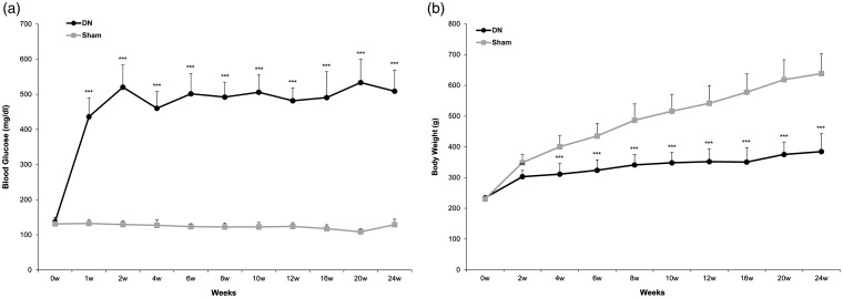
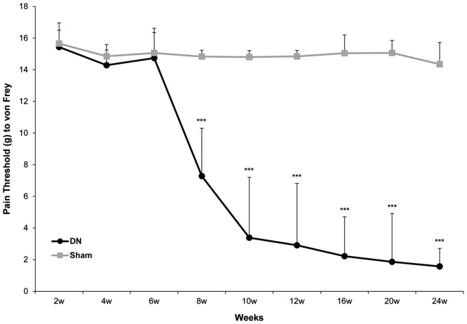
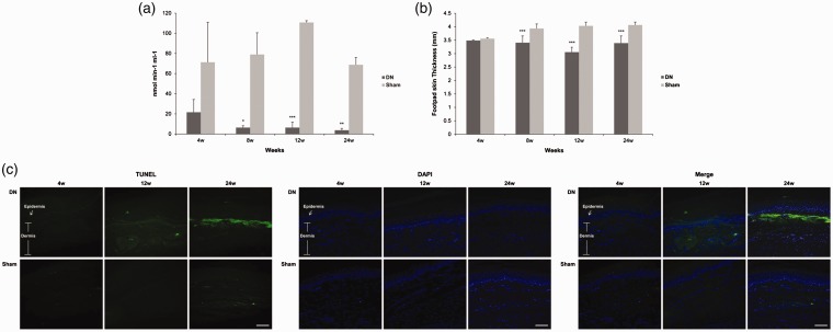
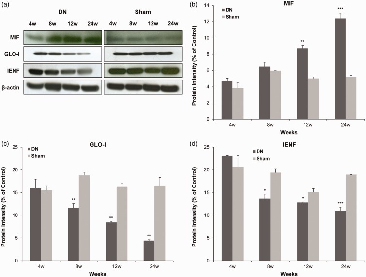
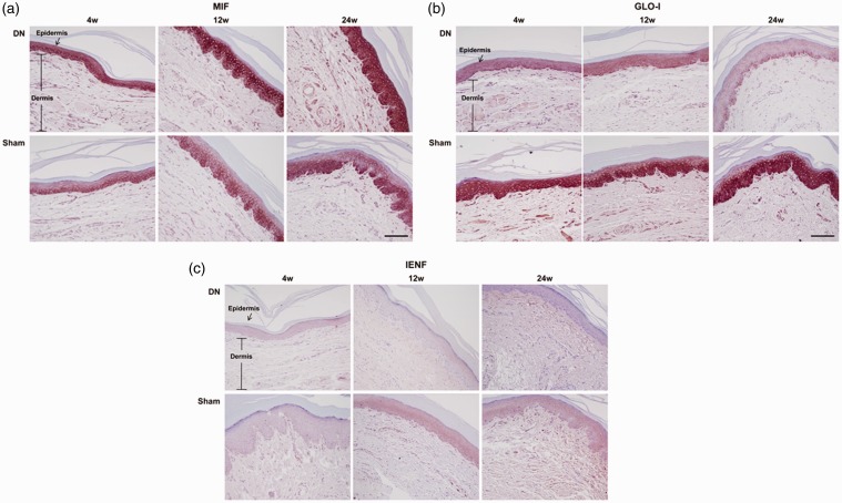
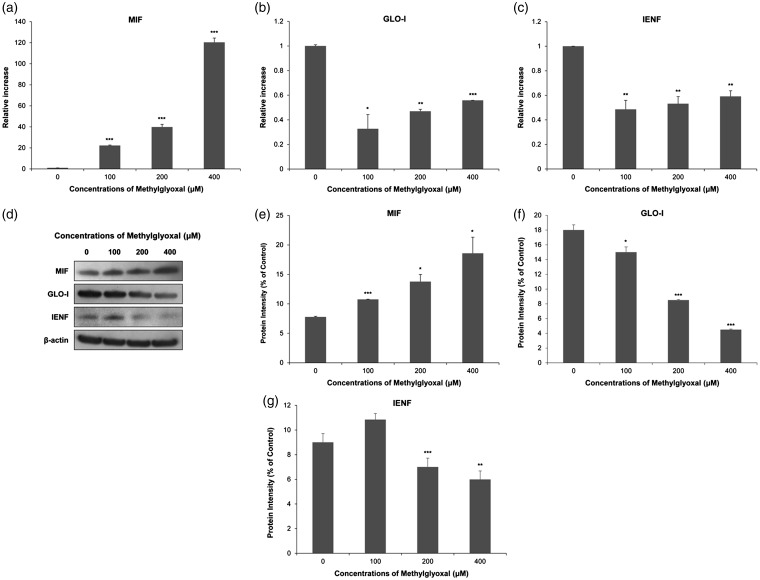
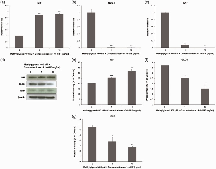
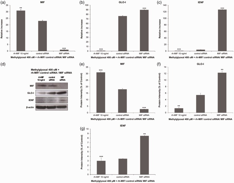
Background Diabetic neuropathy originating in distal lower extremities is associated with pain early in the disease course, overwhelming in the feet. However, the pathogenesis of diabetic neuropathy remains unclear. Macrophage migration inhibitory factor has been implicated in the onset of neuropathic pain and the development of diabetes. Objective of this study was to observe pain syndromes elicited in the footpad of diabetic neuropathy rat model and to assess the contributory role of migration inhibitory factor in the pathogenesis of diabetic neuropathy. Methods Diabetic neuropathy was made in Sprague Dawley rats by streptozotocin. Pain threshold was evaluated using von Frey monofilaments for 24 weeks. On comparable experiment time after streptozotocin injection, all footpads were prepared for following procedures; glutathione assay, terminal deoxynucleotidyl transferase-mediated biotinylated UTP nick end labeling staining, immunohistochemistry staining, real-time reverse transcription polymerase chain reaction, and Western blot. Additionally, human HaCaT skin keratinocytes were treated with methylglyoxal, transfected with migration inhibitory factor/control small interfering RNA, and prepared for real-time reverse transcription polymerase chain reaction and Western blot. Results As compared to sham group, pain threshold was significantly reduced in diabetic neuropathy group, and glutathione was decreased in footpad skin, simultaneously, cell death was increased. Over-expression of migration inhibitory factor, accompanied by low expression of glyoxalase-I and intraepidermal nerve fibers, was shown on the footpad skin lesions of diabetic neuropathy. But, there was no significance in expression of neurotransmitters and inflammatory mediators such as transient receptor potential vanilloid 1, mas-related G protein coupled receptor D, nuclear factor kappa B, tumor necrosis factor-alpha, and interleukin-6 between diabetic neuropathy group and sham group. Intriguingly, small interfering RNA-transfected knockdown of the migration inhibitory factor gene in methylglyoxal-treated skin keratinocytes increased expression of glyoxalase-I and intraepidermal nerve fibers in comparison with control small interfering RNA-transfected cells, which was decreased by induction of methylglyoxal. Conclusions Our findings suggest that migration inhibitory factor can aggravate diabetic neuropathy by suppressing glyoxalase-I and intraepidermal nerve fibers on the footpad skin lesions and provoke pain. Taken together, migration inhibitory factor might offer a pharmacological approach to alleviate pain syndromes in diabetic neuropathy.

Keywords: Diabetic neuropathy; footpad skin; glyoxalase-I; intraepidermal nerve fibers; macrophage migration inhibitory factor; pain.


Figures

Figure 1.
Figure 1.
(a) Blood glucose level and (b) body weight changes after streptozotocin (STZ) or saline treatment in Sprague Dawley rats. Experimental diabetic neuropathy (DN) was induced in male Sprague Dawley rats weighing 250 to 300g by a single intraperitoneal injection of 70 mg/kg STZ to achieve a maximal induced diabetic ratio and maintain a stable and chronic hyperglycemic state. Diabetes was confirmed by a persistently elevated blood glucose level greater than 270 mg/dL. Blood glucose level and body weight were checked for 24 weeks. All values are expressed as mean ± standard deviation (SD) (n= 6 per group). **P<0.01, ***P<0.005 compared to those in the sham group.
Figure 2.
Figure 2.
Behavior phenotype of mechanical sensitivity in a diabetic neuropathy (DN) rat model. Mechanical threshold was evaluated weekly using a set of von Frey monofilaments (0.4, 0.6, 1, 2, 4, 6, 8, and 15 g) applied to the plantar surface of the hind paw. Beginning with 2-g monofilament, the next smaller filament was used if there was a negative response or the next larger gram filament was used if there was a positive response. All data are expressed as mean ± standard deviation (SD) (n= 6 per group). ***P<0.005 compared to that in the sham group.
Figure 3.
Figure 3.
Changes of (a) glutathione (GSH) level, (b) footpad skin thickness, and (c) terminal deoxynucleotidyl transferase biotin-dUTP nick-end labeling (TUNEL, green), 4′, 6-diamidino-2-phenylindole (blue) staining in footpad skin lesions from a diabetic neuropathy rat model. Footpad thickness was measured using a dial caliper. Footpad skin tissues were excised, and GSH levels were measured using GSH assay kit. Some footpads were fixed in 10% formalin and embedded in paraffin. De-paraffinized sections (4 μm thick) were stained for TUNEL assay. Values are shown as mean ± standard deviation (SD) (n= 6 per group). Sections were observed with magnification of ×200, scale bar (white) = 60 μm. *P< 0.05, **P<0.01, ***P<0.005 compared to that in the sham group.
Figure 4.
Figure 4.
Expressions levels of macrophage migration inhibitory factor (MIF), glyoxalase I (GLO-I), and intraepidermal nerve fibers (IENF) mRNAs in footpad skin lesions in a diabetic neuropathy (DN) rat model. Total RNA (10 μg) obtained from each footpad tissue sample was reverse transcribed into cDNA. Real-time polymerase chain reaction was performed with a SYBR Green assay system. Relative gene expression levels normalized to that of β-actin was determined with 2−ΔΔCT method. All columnar values are expressed as mean ± standard deviation (SD) (n= 6 per group). *P<0.05, **P<0.01, ***P<0.005 compared to that in the sham group.
Figure 5.
Figure 5.
Protein expression levels of macrophage migration inhibitory factor (MIF), glyoxalase I (GLO-I), and intraepidermal nerve fibers (IENF) in footpad skin lesions in a diabetic neuropathy (DN) rat model. Protein (20 μg) was obtained from each footpad tissue sample. Protein levels were examined by Western blot. As a control for Western blot analysis, the level of β-actin was determined using an antibody against β-actin. All columnar values are expressed as mean ± standard deviation (SD) (n= 6 per group). *P<0.05, **P<0.01, ***P<0.005 compared to that in the sham group.
Figure 6.
Figure 6.
Histological expressions of macrophage migration inhibitory factor (MIF), glyoxalase I (GLO-I), and intraepidermal nerve fibers (IENF) in footpad skin lesions in a diabetic neuropathy (DN) rat model. Footpads were fixed in 10% formalin and embedded in paraffin. De-paraffinized sections (4 μm thick) were prepared for immunohistochemistry staining. They were examined under a light microscope to assess histological changes. Sections were observed at magnification of ×200. Scale bar (black) = 60 μm. A representative picture from three independent immunohistochemistry staining experiments is shown.
Figure 7.
Figure 7.
Macrophage migration inhibitory factor (MIF), glyoxalase I (GLO-I), and intraepidermal nerve fibers (IENF) expression level changes in human HaCaT keratinocytes after being exposed to various concentrations of methylglyoxal (MG). Expression levels of MIF, GLO-I, and IENF mRNAs and proteins were measured by real-time reverse transcription polymerase chain reaction (a) to (c) and Western blot (d) to (g), respectively. Real-time PCR analysis was performed with a SYBR Green assay system. Relative gene expression level normalized to β-actin was calculated with 2−△△CT method. As a control for Western blot analysis, the level of β-actin was determined using an antibody against β-actin. All results are presented as mean ± SD of three independent experiments. *P< 0.05, **P<0.01, ***P<0.005 compared to that in the negative control group.
Figure 8.
Figure 8.
Macrophage migration inhibitory factor (MIF), glyoxalase I (GLO-I), and intraepidermal nerve fibers (IENF) expression level changes in human HaCaT keratinocytes after being exposed to recombinant human (rh)-MIF in the presence of 400 μm MG. Expression levels of MIF, GLO-I, and IENF mRNAs and proteins were measured by real-time reverse transcription polymerase chain reaction (a) to (c) and Western blot (d) to (g), respectively. Real-time PCR analysis was performed with a SYBR Green assay system. Relative gene expression level normalized to β-actin was calculated with 2−△△CT method. As a control for Western blot analysis, the level of β-actin was determined using an antibody against β-actin. All results are presented as mean ± SD of three independent experiments. *P<0.05, **P<0.01, ***P<0.005 compared to that in the negative control group.
Figure 9.
Figure 9.
Effects of macrophage migration inhibitory factor (MIF) small interfering RNA (siRNA) in the presence of 400 μm methylglyoxal (MG) in human HaCaT keratinocytes. Cells were transfected with MIF siRNA or a control siRNA as well as recombinant human (rh)-MIF before stimulation with MG. Expression levels of MIF, glyoxalase I (GLO-I), and intraepidermal nerve fibers (IENF) mRNAs and proteins were measured by real-time reverse transcription polymerase chain reaction (a) to (c) and Western blot (d) to (g), respectively. Real-time PCR analysis was performed with a SYBR Green assay system. Relative gene expression levels normalized to β-actin was calculated with 2−△△CT method. As a control for Western blot analysis, the level of β-actin was determined using an antibody against β-actin. All results are presented as mean ± SD of three independent experiments. ++P< 0.01, +++P<0.005 compared to that in the group transfected with control siRNA.

Similar articles

Cited by

References

    1. Boulton AJM Vinik AI Arezzo JC, Bril V, Feldman EL, Freeman R, Malik RA, Maser RE, Sosenko JM and Ziegler D. Diabetic neuropathies: a statement by the American Diabetes Association. Diabetes Care 2005; 28: 956–962. - PubMed
    1. Britland ST Young RJ Sharma AK and Clarke, BF. Association of painful and painless diabetic polyneuropathy with different patterns of nerve fiber degeneration and regeneration. Diabetes 1990; 39: 898–908. - PubMed
    1. Boric M, Skopljanac I, Ferhatovic L, Jelicic Kadic A, Banozic A and Puljak L. Reduced epidermal thickness, nerve degeneration and increased pain-related behavior in rats with diabetes type 1 and 2. J Chem Neuroanat 2013; 53: 33–40. - PubMed
    1. Lauria G, Lombardi R, Borgna M, Penza P, Bianchi R, Savino C, Canta A, Nicolini G, Marmiroli P and Cavaletti G. Intraepidermal nerve fiber density in rat foot pad: neuropathologic-neurophysiologic correlation. J Peripher Nerv Syst 2005; 10: 202–208. - PubMed
    1. Chu CC, Wang SD, Chiang JS and Ho ST. Generalized depletion of free nerve endings and decrease of cutaneous nervous innervation in streptozotocin-induced painful and painless diabetic rats. J Chin Med Assoc 2012; 75: 314–321. - PubMed

Publication types

MeSH terms