. 2018 Jun 22;123(1):73-85.
doi: 10.1161/CIRCRESAHA.118.312589. Epub 2018 Apr 24.

Topological Arrangement of Cardiac Fibroblasts Regulates Cellular Plasticity

Affiliations

Topological Arrangement of Cardiac Fibroblasts Regulates Cellular Plasticity

Jingyi Yu et al. Circ Res. .

Abstract

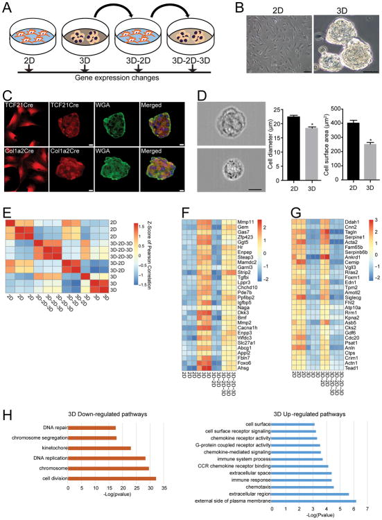
Rationale: Cardiac fibroblasts do not form a syncytium but reside in the interstitium between myocytes. This topological relationship between fibroblasts and myocytes is maintained throughout postnatal life until an acute myocardial injury occurs, when fibroblasts are recruited to, proliferate and aggregate in the region of myocyte necrosis. The accumulation or aggregation of fibroblasts in the area of injury thus represents a unique event in the life cycle of the fibroblast, but little is known about how changes in the topological arrangement of fibroblasts after cardiac injury affect fibroblast function.

Objective: The objective of the study was to investigate how changes in topological states of cardiac fibroblasts (such as after cardiac injury) affect cellular phenotype.

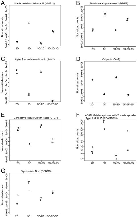
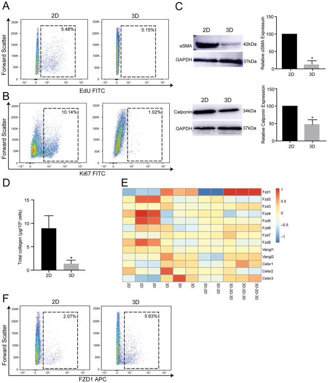
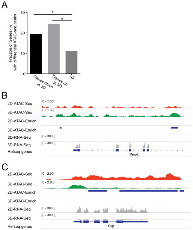
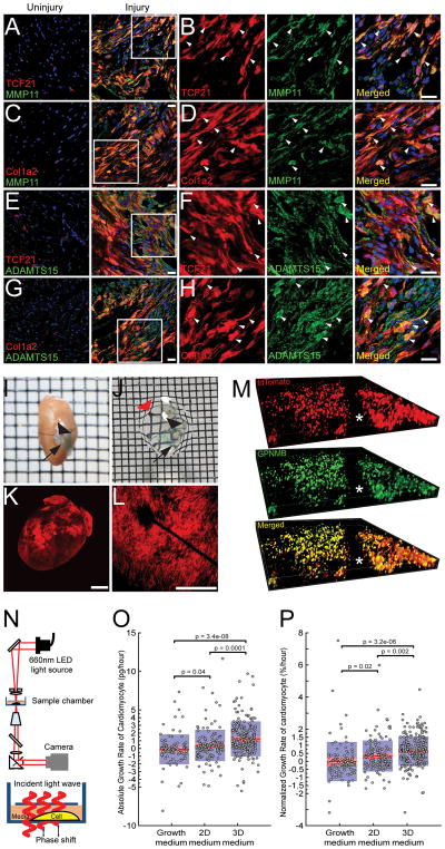
Methods and results: Using 2 and 3-dimensional (2D versus 3D) culture conditions, we show that simple aggregation of cardiac fibroblasts is sufficient by itself to induce genome-wide changes in gene expression and chromatin remodeling. Remarkably, gene expression changes are reversible after the transition from a 3D back to 2D state demonstrating a topological regulation of cellular plasticity. Genes induced by fibroblast aggregation are strongly associated and predictive of adverse cardiac outcomes and remodeling in mouse models of cardiac hypertrophy and failure. Using solvent-based tissue clearing techniques to create optically transparent cardiac scar tissue, we show that fibroblasts in the region of dense scar tissue express markers that are induced by fibroblasts in the 3D conformation. Finally, using live cell interferometry, a quantitative phase microscopy technique to detect absolute changes in single cell biomass, we demonstrate that conditioned medium collected from fibroblasts in 3D conformation compared with that from a 2D state significantly increases cardiomyocyte cell hypertrophy.

Conclusions: Taken together, these findings demonstrate that simple topological changes in cardiac fibroblast organization are sufficient to induce chromatin remodeling and global changes in gene expression with potential functional consequences for the healing heart.

Keywords: cell biology; fibroblasts; fibrosis; hypertrophy; interferometry.


Figures

Figure 1
Figure 1. Cardiac fibroblasts exhibit dynamic changes in gene expression in different topological states
(A) Schematic of how fibroblasts were transitioned from a 2D to 3D state and then back to 2D and 3D respectively. For each topological state, fibroblasts were harvested for RNA-seq. (B) Bright phase image of cardiac fibroblasts in 2D and 3D (Scale bar: 50μm) (C) Pure population of genetically labeled (tdTomato) fibroblasts isolated by flow cytometry from hearts of TCF21MerCreMer:R26Rtdtomato or Col1a2CreERT:R26Rtdtomato mice were subjected to sphere formation (3D) and spheres stained with wheat germ agglutinin (WGA), that stains cell membranes (Scale bar: 20μm). (D) Cardiac fibroblasts in 2D or 3D states dissociated and subjected to image flow cytometry showing representative image of fibroblast from 2D or 3D state (3000 cells imaged in each group, scale bar:10μm) and corresponding mean diameter and surface area of fibroblasts in 2D or 3D states (*p<0.001, mean ± S.E.M., n=3) (E) Heat map demonstrating clustering of sample correlations of fibroblasts (shown by Z scores) in different topological states (F,G) Heat map comparing (F) expression of the most upregulated 3D genes in different topological states and (G) 3D downregulated genes in different topological states (H) GO analysis showing cellular pathways most affected by genes upregulated or downregulated in 2D/3D states.
Figure 2
Figure 2. Dynamic changes in expression of myofibroblast and extracellular matrix genes between 2D and 3D cardiac fibroblast states
(A, B) normalized gene counts on RNA-seq demonstrating rapid changes in gene expression of (A) MMP11 (B) MMP2, (C) Acta2 (D) Calponin (E) Connective tissue growth factor (CTGF) (F) ADAMTS15 and (G) GPNMB in cardiac fibroblasts in different topological states.
Figure 3
Figure 3. Changes in fibroblast phenotype in 3D versus 2D topological state
(A, B) Flow cytometry to determine fraction of proliferating fibroblasts in 2D and 3D states by (A) EdU uptake (5.48±1.4% in 2D versus 0.15±0.05% in 3D, mean ± S.E.M, p<0.05, n=3) or (B) Ki67 expression (10.14±3.0% in 2D versus 1.02±0.01% in 3D, mean ± S.E.M, p<0.05, n=3). (C) Western blotting and quantitative densitometry of expression of Alpha smooth muscle actin and calponin expression by cardiac fibroblasts in 2D or 3D states (mean ± S.E.M, *p<0.001, n=3). (D) Estimation of total collagen content of cardiac fibroblasts in 2D or 3D state (8.40±2.8μg/106 cells in 3D versus 1.32±0.71/106 cells in 2D, mean ± S.E.M, *p<0.05, n=3). (E) Heat map demonstrating expression of members of the Frizzled, Vangl and Celsr family in different topological states of cardiac fibroblasts. (F) Flow cytometry demonstrating Fzd1 expression in 3D versus 2D cardiac fibroblasts (2.07±0.33% in 2D versus 5.63±0.24% in 3D, mean ± S.E.M, p<0.05, n=3).
Figure 4
Figure 4. Chromatin changes underlie altered gene expression of fibroblasts in 3D versus 2D states
(A) ATAC seq performed to demonstrate fraction of genes demonstrating differential ATAC-seq peaks in either 2D or 3D cardiac fibroblast states (B, C) ATAC-seq peaks and RNA-seq showing expression of (B) MMP2 and (C) CTGF in 2D and 3D states demonstrating differential ATAC-seq peaks in loci of MMP2 and CTGF genes (numbers listed refer to scales of enrichment).
Figure 5
Figure 5. Genes enriched in 3D fibroblast states show significant correlation with indices of adverse ventricular modeling in HMDP studies following isoproterenol infusion
(A) Correlation heat map (yellow: positive and blue: negative correlation) of top 15 differentially upregulated genes in 3D/2D states versus clinical traits of left ventricular dimensions, heart mass, plasma glucose and heart rate following infusion of isoproterenol (B) Individual gene contribution to eigengene signatures PC1 and PC2 using transcripts enriched in 3D states. (C–H) Correlation of both eigengene signatures against cardiac and non-cardiac traits with significant correlation between both eigengenes and (C, D) LVID at end diastole (E, F) LVID at end systole and (G, H) total heart mass with no significant correlation between either eigengene and (I–J) heart rate and (K, L) plasma glucose (LVID: left ventricular internal diameter; bicor: bicorrelation coefficient).
Figure 6
Figure 6. Genes enriched in 3D fibroblasts are expressed in vivo in regions of fibroblast aggregation after heart injury and affect cardiomyocyte hypertrophy
(A–D) Immunofluorescent staining for MMP11 on uninjured and cryo-injured hearts of (A, B) TCF21MerCreMer:R26Rtdtomato and (C, D) Col1a2CreERT:R26Rtdtomato mice (B, D) area of injury shown in higher magnification demonstrating tdTomato labeled fibroblasts expressing MMP11 (arrows). (E–H) Immunofluorescent staining for ADAMTS15 on uninjured and cryo-injured hearts of (E, F) TCF21MerCreMer:R26Rtdtomato and (G, H) area of injury shown in higher magnification demonstrating tdTomato labeled fibroblasts expressing ADAMTS15 (arrows) (Scale bars: 20μm). (I, J) Cryo-injured heart of Col1a2CreERT:R26Rtdtomato mouse (I) prior to and (J) following optical clearing (arrowhead points to suture for identifying injured region, arrow points to green dye to identify area adjacent to injury; note the wire mesh on which the heart lies is now visible through the transparent heart; red arrow) (K) tdTomato fluorescence observed on cryo-injured Col1a2CreERT:R26Rtdtomato heart and (L) confocal image through area of injury showing intense tdTomato fluorescence (Scale bar: 500μm) (M) Immunofluorescent staining for GPNMB on optically cleared Col1a2CreERT:R26Rtdtomato heart after injury. The entire depth of the scar was imaged with a confocal microscope and sequential Z stack images demonstrating distribution of tdTomato (red), GPNMB (green) and merged (yellow) image demonstrating distribution of fluorophores across the depth of the scar (asterisk corresponds to position of suture). (N) Set up of live cell interferometry with phase shift of light being a read out for changes in cell biomass (O) Absolute and (P) normalized single cell cardiomyocyte (NRVM) biomass accumulation rates determined by serial measurements with interference microscopy over 48 hours following treatment of NRVM with growth medium (non-conditioned) or conditioned medium from fibroblasts in 2D or 3D state (each circle represents a single cardiomyocyte; Number of single cardiomyocytes tracked: 103 for growth medium, 142 for 2D medium and 231 for 3D medium).

Comment in

References

    1. Manner J, Perez-Pomares JM, Macias D, Munoz-Chapuli R. The origin, formation and developmental significance of the epicardium: a review. Cells Tissues Organs. 2001;169:89–103. - PubMed
    1. Christia P, Bujak M, Gonzalez-Quesada C, Chen W, Dobaczewski M, Reddy A, Frangogiannis NG. Systematic characterization of myocardial inflammation, repair, and remodeling in a mouse model of reperfused myocardial infarction. J Histochem Cytochem. 2013;61:555–70. - PMC - PubMed
    1. Camelliti P, Borg TK, Kohl P. Structural and functional characterisation of cardiac fibroblasts. Cardiovasc Res. 2005;65:40–51. - PubMed
    1. Shoval H, Karsch-Bluman A, Brill-Karniely Y, Stern T, Zamir G, Hubert A, Benny O. Tumor cells and their crosstalk with endothelial cells in 3D spheroids. Scientific Reports. 2017;7:10428. - PMC - PubMed
    1. Pillai IC, Li S, Romay M, Lam L, Lu Y, Huang J, Dillard N, Zemanova M, Rubbi L, Wang Y, Lee J, Xia M, Liang O, Xie YH, Pellegrini M, Lusis AJ, Deb A. Cardiac Fibroblasts Adopt Osteogenic Fates and Can Be Targeted to Attenuate Pathological Heart Calcification. Cell stem cell. 2017;20:218–232e5. - PMC - PubMed

Publication types

Substances