. 2018 Jun 15;293(24):9461-9472.
doi: 10.1074/jbc.RA117.001658. Epub 2018 Apr 24.

The compound millepachine and its derivatives inhibit tubulin polymerization by irreversibly binding to the colchicine-binding site in β-tubulin

Affiliations

The compound millepachine and its derivatives inhibit tubulin polymerization by irreversibly binding to the colchicine-binding site in β-tubulin

Jianhong Yang et al. J Biol Chem. .

Abstract

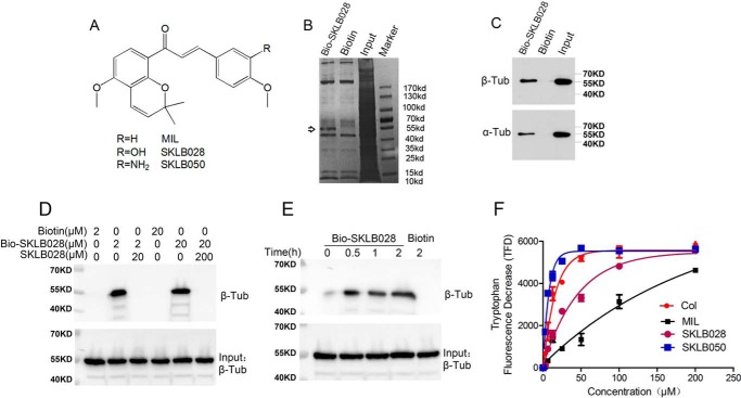
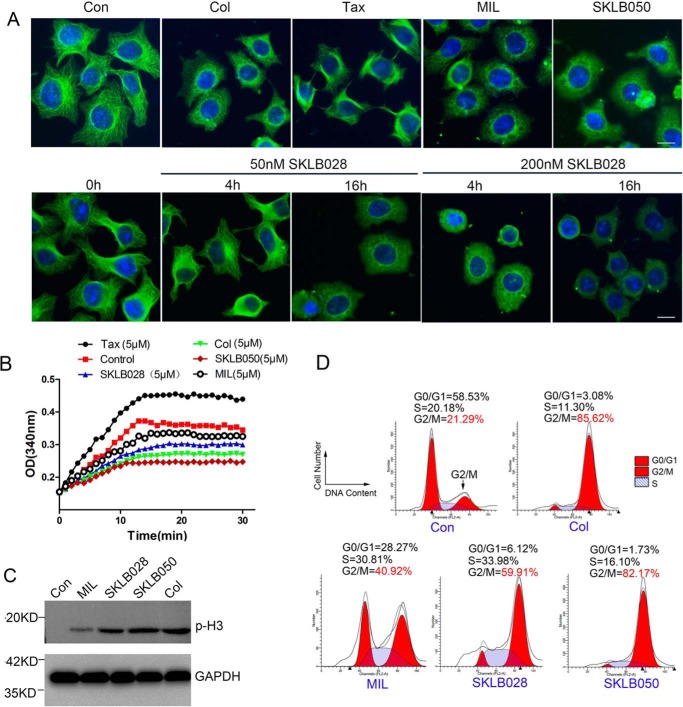
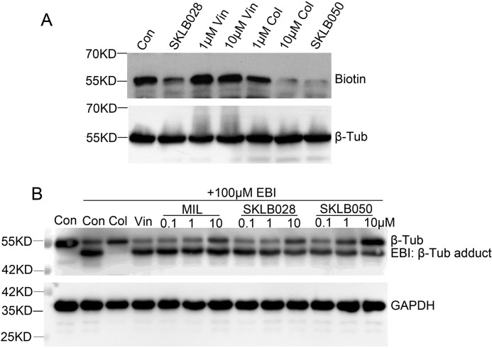
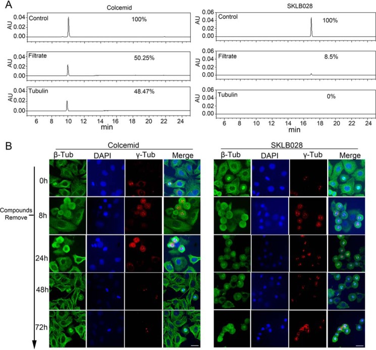
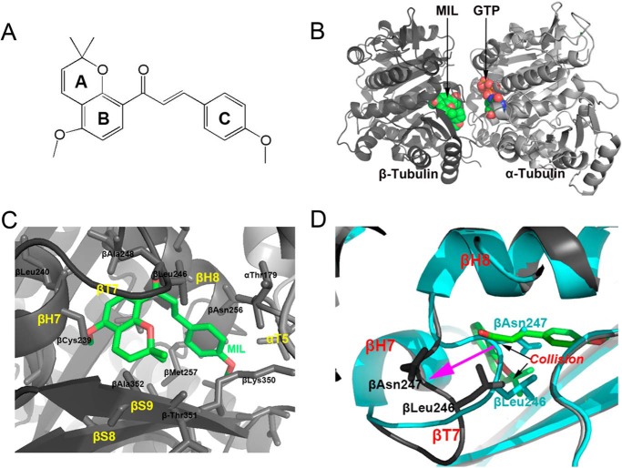
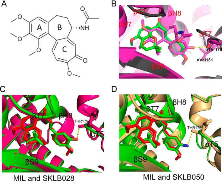
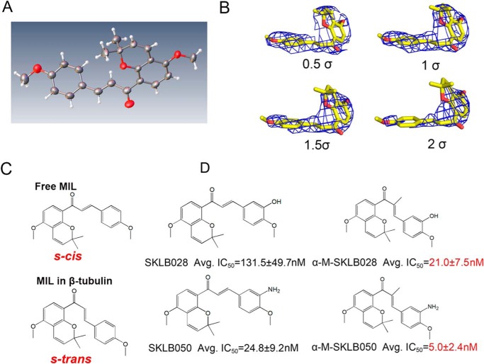
Inhibitors that bind to the paclitaxel- or vinblastine-binding sites of tubulin have been part of the pharmacopoeia of anticancer therapy for decades. However, tubulin inhibitors that bind to the colchicine-binding site are not used in clinical cancer therapy, because of their low therapeutic index. To address multidrug resistance to many conventional tubulin-binding agents, numerous efforts have attempted to clinically develop inhibitors that bind the colchicine-binding site. Previously, we have found that millepachine (MIL), a natural chalcone-type small molecule extracted from the plant Millettia pachycarpa, and its two derivatives (MDs) SKLB028 and SKLB050 have potential antitumor activities both in vitro and in vivo However, their cellular targets and mechanisms are unclear. Here, biochemical and cellular experiments revealed that the MDs directly and irreversibly bind β-tubulin. X-ray crystallography of the tubulin-MD structures disclosed that the MDs bind at the tubulin intradimer interface and to the same site as colchicine and that their binding mode is similar to that of colchicine. Of note, MDs inhibited tubulin polymerization and caused G2/M cell-cycle arrest. Comprehensive analysis further revealed that free MIL exhibits an s-cis conformation, whereas MIL in the colchicine-binding site in tubulin adopts an s-trans conformation. Moreover, introducing an α-methyl to MDs to increase the proportion of s-trans conformations augmented MDs' tubulin inhibition activity. Our study uncovers a new class of chalcone-type tubulin inhibitors that bind the colchicine-binding site in β-tubulin and suggests that the s-trans conformation of these compounds may make them more active anticancer agents.

Keywords: Chalcone; Colchicine; Millepachine; Tubulin; X-ray crystallography; cancer; cancer prevention; drug resistance; microtubule; natural product; s-trans conformation; tubulin.


Conflict of interest statement

The authors declare that they have no conflicts of interest with the contents of this article

Figures

Figure 1.
Figure 1.
MIL directly targets β-tubulin. A, chemical structures of MIL. B, HepG2 cell lysates were incubated with Bio-SKLB028 or biotin, followed by pulldown with streptavidin–agarose. The precipitates were resolved by SDS-PAGE, and the gel was stained with Coomassie staining. C, HepG2 cell lysates were incubated with Bio-SKLB028 or biotin, followed by pulldown with streptavidin–agarose. The precipitates were detected by Western blotting by α- and β-tubulin. D, porcine brain tubulin (1 μm) were preincubated with or without indicated concentrations of SKLB028 for 2 h before incubation with lower concentration of Bio-SKLB028 for another 2 h, and different concentrations of biotin were incubated with tubulin for 2 h as control groups. Then all the samples were pulled down by streptavidin–agarose beads, and the eluted samples were bolted for β-tubulin. Also, total protein for each sample was detected by Western blotting for β-tubulin as a loading control. E, porcine brain tubulin (1 μm) was incubated with Bio-SKLB028 for different times and then pulled down by a streptavidin–agarose beads, and the eluted samples were bolted for β-tubulin. Also, total protein for each sample was detected by Western blotting for β-tubulin as a loading control. F, tryptophan-based binding assay to detect the Kd value of indicated compounds binding to tubulin. The indicated compounds at different concentrations (3.125, 6.25, 12.5, 25, 50, 100, and 200 μm) were incubated with tubulin for 30 min and then monitored at 295 nm (excitation) and 335 nm (emission) using Biotech Gen5 spectrophotometer. The dissociation constants were calculated from fitting curves of decreasing fluorescence using the GraphPad Prism software. Col, colchicine; Tub, tubulin; Col, colchicine.
Figure 2.
Figure 2.
MDs inhibit tubulin polymerization and cause G2/M phase cell-cycle arrest. A, HCT-8/V cells were treated with 1 μm MIL, 50 nm SKLB050, 50 nm colchicine, or 50 nm paclitaxel for 16 h or treated with 50 nm or 200 nm SKLB028 for 4 or 16 h. Then tubulin morphology was detected by immunofluorescence with anti-β-tubulin and DAPI staining. Bar, 10 μm. B, indicated concentrations of compounds were co-incubated with tubulin (3 mg/ml) at 37 °C. Absorbance at 340 nm was detected every 1 min for 30 min. C, HCT-8/V cells were incubated with 1 μm MIL, 50 nm SKLB050, 50 nm colchicine, or 100 nm SKLB028 for 16 h; then cell lysates were collected and subjected to Western blotting for p-H3 detection. GAPDH was used as a loading control. D, HCT-8/V cells were incubated with 1 μm MIL, 50 nm SKLB050, 50 nm colchicine, and 100 nm for 16 h; subjected to PI staining; and analyzed by a flow cytometer for cell-cycle analysis. Col, colchicine; Con, control; Tax, paclitaxel.
Figure 3.
Figure 3.
MDs bind to colchicine site of β-tubulin. A, porcine brain tubulin (1 μm) were treated with 1 μm SKLB028, 1 μm vinblastine, 10 μm vinblastine, 1 μm colchicine, 10 μm colchicine, or 1 μm SKLB050 for 1 h before treated with 1 μm Bio-SKLB028 for another 2 h. After totally wash of unbounded compounds and cross-linked by a UV hybridization incubator for 10 min, the samples were blotted for biotin. Total proteins were blotted for β-tubulin as a loading control. B, HCT-8/V cells were incubated with or without indicated concentrations of MDs for 2 h, and then 100 μm EBI was added to cells and incubated for another 2 h. Total protein was lysed with RIPA lysis buffer and subjected to Western blotting analysis for β-tubulin and GAPDH. Vin, vinblastine; Col, colchicine; Con, control; Tub, tubulin.
Figure 4.
Figure 4.
MDs bind irreversibly to β-tubulin. A, tubulin (3 mg/ml) incubated with 30 μm colcemid or 30 μm SKLB028 for 1 h. Then tubulin part and filtrate part were separated with an ultrafiltration method. Compounds in tubulin part and filtrate part were detected by HPLC. Colcemid (30 μm) or SKLB028 (30 μm) dissolved in 200 μl of CH3OH were also detected by HPLC as controls. This experiment was repeated three times. B, high concentrations (10 μm) of SKLB028 or colcemid were co-incubated with HCT-8/V cells for 8 h, and then compounds were extensively washed for three times. The cells were further cultured to 72 h. At 0-, 8-, 24-, 48-, and 72-h points, tubulin morphology was analyzed by immunofluorescence. Bar, 20 μm. Tub, tubulin; AU, absorbance unit.
Figure 5.
Figure 5.
Structure of the MIL–T2R–TTL complex. A, chemical structure of MIL. B, overall view of the complex formed between αβ-tubulin and MIL. C, close-up views of the interaction network observed between MIL (green sticks) and tubulin (black cartoon). Interacting residues of tubulin are shown in stick representation. Oxygen atoms are colored red and blue. D, superimposition of the MIL–T2R–TTL (cyan cartoon) and apo-T2R–TTL (black cartoon) structures. MIL is in green stick representation. Black arrows highlight regions of steric clashes between tubulin and MIL, purple arrow shows the large main chain changes between apo- from MIL complex.
Figure 6.
Figure 6.
Comparison the binding modes of MIL– and colchicine–, MIL– and SKLB028–, MIL– and SKLB050–T2R–TTL complex. A, chemical structure of colchicine. B, superimposition of the MIL–T2R–TTL (gray cartoons) and colchicine–T2R–TTL (purple cartoons) structures. MIL and colchicine are in green and purple stick representation, respectively. C, superimposition of the MIL–T2R–TTL (green cartoons) and SKLB028–T2R–TTL (purple cartoons) structures. MIL and SKLB028 are in red and green stick representation, respectively. D, superimposition of the MIL–T2R–TTL (green cartoons) and SKLB050–T2R–TTL (pale yellow cartoons) structures. MIL and SKLB050 are in red and green stick representation, respectively.
Figure 7.
Figure 7.
Chalcones with s-trans conformation are more active. A, X-ray structure of free MIL. B, stereo views of MIL in tubulin with the final refined 2FoFc electron density contoured at 0.5, 1, 1.5, and 2 σ. C, structural formula of free MIL (s-cis) and MIL (s-trans) in tubulin complex. D, chemical structures of MDs and their average IC50s on five cancer cells (A2780S, A2780/T, HCT-8, HCT-8/V, and HCT-8/T; original data shown in Table S1).

Similar articles

Cited by

References

    1. Perez, E. A. (2009) Microtubule inhibitors: Differentiating tubulin-inhibiting agents based on mechanisms of action, clinical activity, and resistance. Mol. Cancer Ther. 8, 2086–2095 10.1158/1535-7163.MCT-09-0366 - DOI - PubMed
    1. Jordan, M. A., and Kamath, K. (2007) How do microtubule-targeted drugs work? An overview. Curr. Cancer Drug Targets 7, 730–742 10.2174/156800907783220417 - DOI - PubMed
    1. Bhalla, K. N. (2003) Microtubule-targeted anticancer agents and apoptosis. Oncogene 22, 9075–9086 10.1038/sj.onc.1207233 - DOI - PubMed
    1. Michaud, L. B. (2009) The epothilones: how pharmacology relates to clinical utility. Ann. Pharmacother. 43, 1294–1309 10.1345/aph.1M005 - DOI - PubMed
    1. Kim, J. S., Lee, Y. C., Nam, H. T., Li, G., Yun, E. J., Song, K. S., Seo, K. S., Park, J. H., Ahn, J. W., Zee, O., Park, J. I., Yoon, W. H., Lim, K., and Hwang, B. D. (2007) Apicularen A induces cell death through Fas ligand up-regulation and microtubule disruption by tubulin down-regulation in HM7 human colon cancer cells. Clin. Cancer Res. 13, 6509–6517 10.1158/1078-0432.CCR-07-1428 - DOI - PubMed

Publication types

MeSH terms