. 2018 Jul 2;217(7):2503-2518.
doi: 10.1083/jcb.201711037. Epub 2018 Apr 24.

Hierarchical recruitment of ribosomal proteins and assembly factors remodels nucleolar pre-60S ribosomes

Affiliations

Hierarchical recruitment of ribosomal proteins and assembly factors remodels nucleolar pre-60S ribosomes

Stephanie Biedka et al. J Cell Biol. .

Abstract

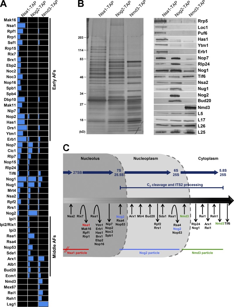
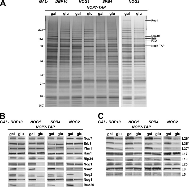
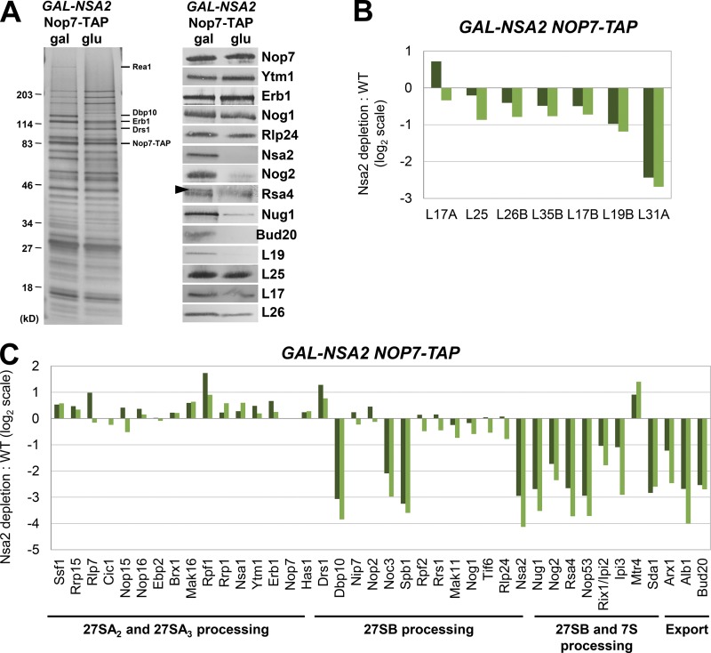
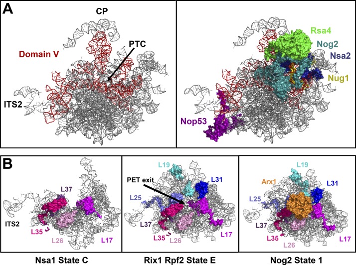
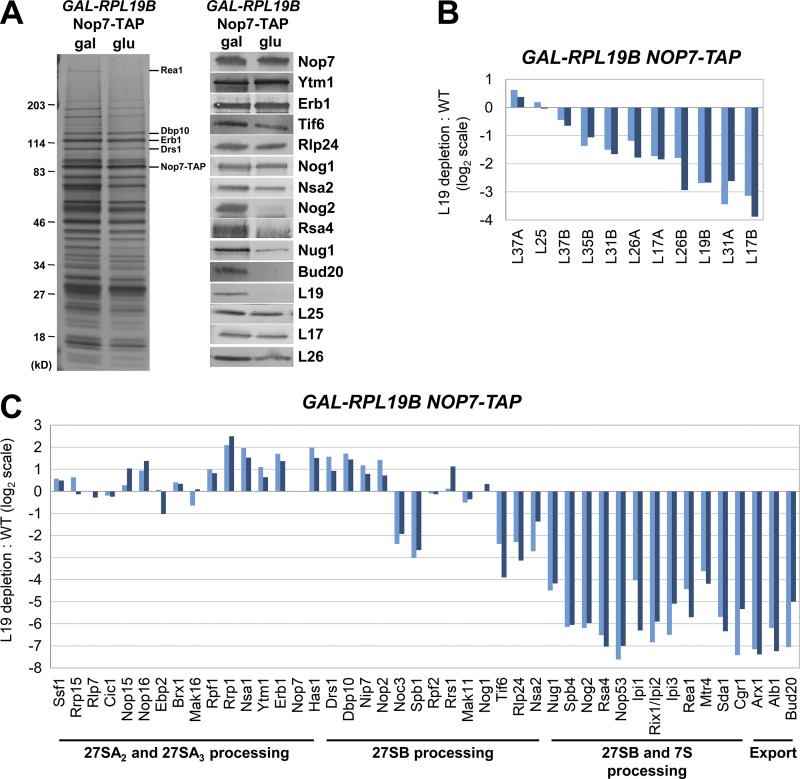
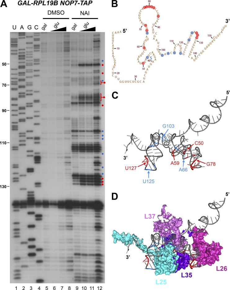
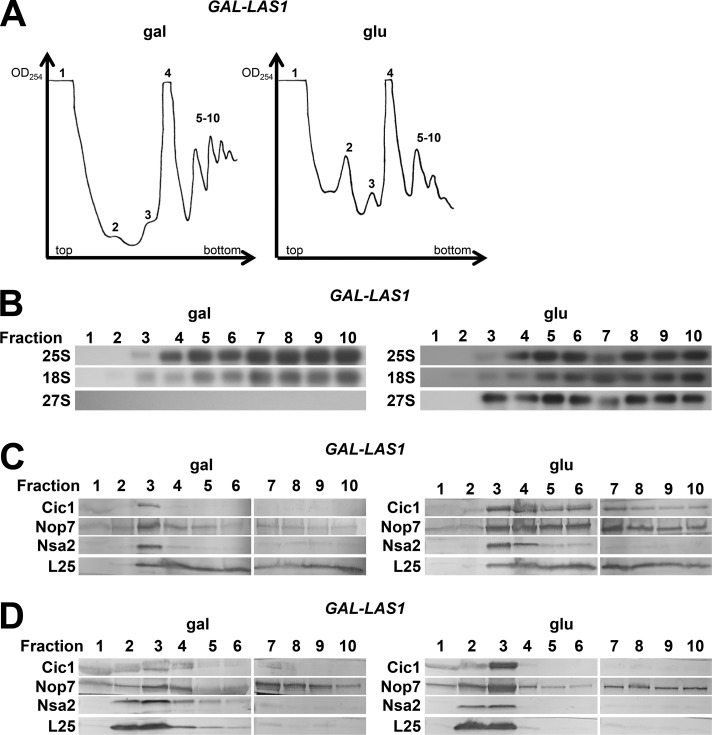
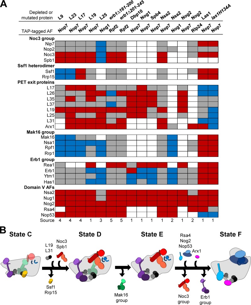
Ribosome biogenesis involves numerous preribosomal RNA (pre-rRNA) processing events to remove internal and external transcribed spacer sequences, ultimately yielding three mature rRNAs. Removal of the internal transcribed spacer 2 spacer RNA is the final step in large subunit pre-rRNA processing and begins with endonucleolytic cleavage at the C2 site of 27SB pre-rRNA. C2 cleavage requires the hierarchical recruitment of 11 ribosomal proteins and 14 ribosome assembly factors. However, the function of these proteins in C2 cleavage remained unclear. In this study, we have performed a detailed analysis of the effects of depleting proteins required for C2 cleavage and interpreted these results using cryo-electron microscopy structures of assembling 60S subunits. This work revealed that these proteins are required for remodeling of several neighborhoods, including two major functional centers of the 60S subunit, suggesting that these remodeling events form a checkpoint leading to C2 cleavage. Interestingly, when C2 cleavage is directly blocked by depleting or inactivating the C2 endonuclease, assembly progresses through all other subsequent steps.


Figures

Figure 1.
Figure 1.
Pre-60S subunit protein composition changes dramatically leading up to and during ITS2 processing. (A and B) Sequential pre-60S subunit assembly intermediates were purified from yeast via TAP-tagged AFs Nsa1, Nog2, or Nmd3. The protein composition of the purified pre-60S subunits was analyzed via mass spectrometry (A) and by SDS-PAGE followed by silver staining and Western blotting (B). (C) A working model for the pathway of 60S subunit assembly. The association and dissociation of AFs with pre-60S subunits are indicated as is the timing of pre-rRNA processing events. The lifetimes of the Nsa1, Nog2, and Nmd3 particles are shown; Nsa1 enters preribosomes during early stages of 60S subunit assembly.
Figure 2.
Figure 2.
Similar changes in pre-60S subunit protein composition occur upon depletion of the B-factors Dbp10, Nog1, Spb4, or Nog2. The TAP-tagged AF Nop7 was used as bait to purify pre-60S subunits from yeast expressing (gal) or depleted of (glu) the B-factors Dbp10, Nog1, Spb4, and Nog2. The preribosome protein composition was analyzed by SDS-PAGE followed by silver staining (A) and Western blotting for AFs (B) and r-proteins (C). Asterisks indicate that the protein being blotted for was tagged and detected with anti-HA antibody.
Figure 3.
Figure 3.
Dramatic changes in pre-60S subunit protein composition are caused by depletion of AF Nsa2. (A–C) Preribosomes containing (gal) or lacking (glu) Nsa2 were purified using TAP-tagged Nop7 as bait. (A) The protein composition of the pre-60S subunits was analyzed by SDS-PAGE followed by silver staining and Western blotting. Rsa4 is indicated by a black arrowhead; the contaminating band is IgG. (B and C) Semiquantitative mass spectrometry (iTRAQ) was used to quantify the relative changes in levels of 60S subunit r-proteins (B) and AFs (C) in the presence and absence of Nsa2. The ratios were normalized to levels of Nop7, and the fold change in log2 scale is shown for two biological replicates. A complete dataset for AFs is shown in Fig. S2.
Figure 4.
Figure 4.
AFs binding to 25S rRNA domain V and r-proteins surrounding the PET exit are present in decreased levels in B mutants. (A) The B-factors Nsa2 and Nog2 and the AFs Rsa4, Nug1, and Nop53 bind to 25S rRNA domain V. (B) As assembly proceeds, the globular bodies of the r-proteins L19, L25, L31, L17, L26, L35, and L37 form a platform surrounding the exit of the PET to which Arx1 can then bind. Structures shown are PDB IDs 6EM1, 6ELZ, and 3JCT.
Figure 5.
Figure 5.
Dramatic changes in pre-60S subunit protein composition are caused by depletion of r-protein L19. (A–C) Preribosomes containing (gal) or lacking (glu) L19 were purified using TAP-tagged Nop7 as bait. (A) The protein composition of pre-60S subunits was analyzed by SDS-PAGE followed by silver staining and Western blotting. (B and C) Semiquantitative mass spectrometry (iTRAQ) was used to quantify the relative changes in levels of 60S subunit r-proteins (B) and AFs (C) in the presence and absence of L19. The ratios were normalized to levels of Nop7, and the fold change in log2 scale is shown for two biological replicates. Both samples of yeast grown in glucose were compared with a single sample grown in galactose. A complete dataset for AFs is shown in Fig. S2.
Figure 6.
Figure 6.
The structure of 5.8S rRNA in preribosomes is altered in the absence of L19. (A) In vivo structure probing of 5.8S rRNA (SHAPE) was conducted by treating cells with NAI and assaying modified nucleotides via primer extension with an oligonucleotide complimentary to the 5′ half of ITS2. Cells were grown in galactose or grown in galactose and shifted to glucose for 16 h. Nucleotides with increased or decreased modification in the absence of L19 are indicated by red and blue circles, respectively. RNA extracted from cells treated with DMSO instead of NAI was used as a control. Increasing amounts of primer extension product from cells shifted to glucose were loaded in lanes 6–8 and 10–12 to achieve loading similar to the galactose lanes (5 and 9). (B and C) Nucleotides modified by NAI are indicated on the secondary (B) and tertiary (C) structures of 5.8S rRNA. (D) The PET exit r-proteins L25, L26, L35, and L37 are clustered on top of the modified nucleotides.
Figure 7.
Figure 7.
Depletion or mutational inactivation of Las1 results in changes in pre-60S subunit protein composition unlike those observed in other B mutants. (A and B) Preribosomes were purified from yeast expressing (gal) or depleted of (glu) Las1 or from yeast expressing las1R129A or las1H134A using Nop7-TAP as bait. The protein composition of the pre-60S subunits was analyzed by SDS-PAGE followed by silver staining (A) and Western blotting (B). Yeast containing pRS315-las1R129A or pRS315-las1H134A were grown in C-leu minimal media.
Figure 8.
Figure 8.
AFs bound to the ITS2 foot are present in cytoplasmic pre-60S subunits in the absence of Las1. (A and B) Pre-60S subunits containing (gal) or lacking (glu) Las1 were purified using TAP-tagged Nmd3 as bait. The protein composition of preribosomes was analyzed by SDS-PAGE followed by silver staining (A) and Western blotting (B). (C) The cellular localization of GFP-tagged Nop7 and Cic1 was monitored in live yeast cells expressing (gal) or depleted of (glu) Las1. Bar, 5 µm. (D) The locations of AFs bound on or proximal to ITS2 in the state E particle. The structure shown is PDB ID 6ELZ.
Figure 9.
Figure 9.
27S pre-rRNA and ITS2 AFs are present in polyribosomes in the absence of Las1. (A) Whole-cell lysates from cells expressing (gal) or depleted of (glu) Las1 were fractionated on a 7–47% sucrose gradient, and 1-ml fractions were collected from the indicated peaks. Fractions 1–4 were collected at the maximum of the indicated peak, and fractions 5–10 were collected continuously beginning with the first polyribosome peak. (B and C) Fractions were analyzed by Northern blotting (B) and Western blotting (C). (D) Whole-cell lysates prepared under polyribosome run-off conditions (omission of cycloheximide and MgCl2) from cells expressing or lacking Las1 were fractionated and analyzed by Western blotting.
Figure 10.
Figure 10.
Six common defects occur in most mutants blocked at C2 cleavage. (A) Comparisons of the data presented in this study with previously published data for mutants blocked at C2 cleavage revealed that there are six groups of proteins commonly affected in these mutants. Increased, decreased, or unchanged levels of proteins are indicated by blue, red, and gray boxes, respectively. White boxes indicate that data were not available. Sources: 1, this study; 2, Lebreton et al., 2008; 3, Ohmayer et al., 2013; 4, Gamalinda et al., 2014; 5, Konikkat et al., 2017. (B) A model of the nucleolar remodeling events affected in B mutants.

References

    1. Ausubel F.M., Brent R., Kingston R.E., Moore D.D., Seidman J.G., Smith J.A., and Struhl K.. 1994. Current protocols in molecular biology. John Wiley & Sons Inc, New York.
    1. Bassler J., Kallas M., Pertschy B., Ulbrich C., Thoms M., and Hurt E.. 2010. The AAA-ATPase Rea1 drives removal of biogenesis factors during multiple stages of 60S ribosome assembly. Mol. Cell. 38:712–721. 10.1016/j.molcel.2010.05.024 - DOI - PMC - PubMed
    1. Ben-Shem A., de Loubresse N.G., Melnikov S., Jenner L., Yusupova G., and Yusupov M.. 2011. The structure of the eukaryotic ribosome at 3.0 Å resolution. Science. 334:1524–1529. 10.1126/science.1212642 - DOI - PubMed
    1. Bradatsch B., Leidig C., Granneman S., Gnädig M., Tollervey D., Böttcher B., Beckmann R., and Hurt E.. 2012. Structure of the pre-60S ribosomal subunit with nuclear export factor Arx1 bound at the exit tunnel. Nat. Struct. Mol. Biol. 19:1234–1241. 10.1038/nsmb.2438 - DOI - PMC - PubMed
    1. Castle C.D., Cassimere E.K., Lee J., and Denicourt C.. 2010. Las1L is a nucleolar protein required for cell proliferation and ribosome biogenesis. Mol. Cell. Biol. 30:4404–4414. 10.1128/MCB.00358-10 - DOI - PMC - PubMed

Publication types

MeSH terms

LinkOut - more resources