. 2018 Apr 24;9(2):e00415-18.
doi: 10.1128/mBio.00415-18.

Novel Insights into Staphylococcus aureus Deep Bone Infections: the Involvement of Osteocytes

Affiliations

Novel Insights into Staphylococcus aureus Deep Bone Infections: the Involvement of Osteocytes

Dongqing Yang et al. mBio. .

Abstract

Periprosthetic joint infection (PJI) is a potentially devastating complication of orthopedic joint replacement surgery. PJI with associated osteomyelitis is particularly problematic and difficult to cure. Whether viable osteocytes, the predominant cell type in mineralized bone tissue, have a role in these infections is not clear, although their involvement might contribute to the difficulty in detecting and clearing PJI. Here, using Staphylococcus aureus, the most common pathogen in PJI, we demonstrate intracellular infection of human-osteocyte-like cells in vitro and S. aureus adaptation by forming quasi-dormant small-colony variants (SCVs). Consistent patterns of host gene expression were observed between in vitro-infected osteocyte-like cultures, an ex vivo human bone infection model, and bone samples obtained from PJI patients. Finally, we confirm S. aureus infection of osteocytes in clinical cases of PJI. Our findings are consistent with osteocyte infection being a feature of human PJI and suggest that this cell type may provide a reservoir for silent or persistent infection. We suggest that elucidating the molecular/cellular mechanism(s) of osteocyte-bacterium interactions will contribute to better understanding of PJI and osteomyelitis, improved pathogen detection, and treatment.IMPORTANCE Periprosthetic joint infections (PJIs) are increasing and are recognized as one of the most common modes of failure of joint replacements. Osteomyelitis arising from PJI is challenging to treat and difficult to cure and increases patient mortality 5-fold. Staphylococcus aureus is the most common pathogen causing PJI. PJI can have subtle symptoms and lie dormant or go undiagnosed for many years, suggesting persistent bacterial infection. Osteocytes, the major bone cell type, reside in bony caves and tunnels, the lacuno-canalicular system. We report here that S. aureus can infect and reside in human osteocytes without causing cell death both experimentally and in bone samples from patients with PJI. We demonstrate that osteocytes respond to infection by the differential regulation of a large number of genes. S. aureus adapts during intracellular infection of osteocytes by adopting the quasi-dormant small-colony variant (SCV) lifestyle, which might contribute to persistent or silent infection. Our findings shed new light on the etiology of PJI and osteomyelitis in general.

Keywords: Staphylococcus aureus; bone infection; osteomyelitis; periprosthetic joint infection; small-colony variant; stress adaptation.


Figures

FIG 1
FIG 1
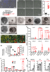
In vitro interaction between primary human-osteocyte-like cells and S. aureus. (A) Viability of osteocyte-like cells determined by DAPI staining of nuclei in the control (Ctrl) and infected (Inf) groups 30 days postinfection. (B) Time-lapse recording (13 to 18 h) of in vitro-differentiated human-osteocyte-like cell movement under S. aureus (strain RN6390; GFP labeled) infection. The sum of plasma membrane movements during the observation period was determined; data shown represent means ± standard errors of the means (SEM) for a pool of 10 cells from each of the Ctrl and Inf groups (P < 0.0001). TEM images of infected osteocyte-like cells showing dying S. aureus cells with compromised cell walls (C), proliferating S. aureus cells with evenly and completely divided cell walls (D), and proliferating S. aureus cells with unevenly and incompletely divided cell walls (E and F). (G) Recovered intracellular S. aureus on days 1, 3, and 5 featured with large and small (red arrowheads) sizes. (H) Quantification of intracellular and extracellular viable S. aureus CFU from days 1 to 5 (left y axis) and the percentage of small colonies recovered from host cells (right y axis) (graphs represent results from 3 independent biological replicates; values are means of results from the pool ± SEM). (I) Relative mRNA levels (normalized to 16S rRNA) of agrA, sarA, asp23, hla, hld, and sigB in recovered small and large colonies (values represent means from 3 independent biological replicates ± SEM). (J) Relative mRNA (normalized to 18S mRNA) levels of CCL5, CXCL1, CXCL8, CXCL9, CXCL10, and CXCL11 at 24 h in osteocyte-like cells under control, killed-S. aureus WCH-SK2 and live-S. aureus WCH-SK2 treatments with an MOI of 100. Data shown are means of normalized expression ± SEM from 4 independent biological replicates. Comparisons (t tests) and P values are indicated. ND, nondetectable. (K) Production of the chemokines RANTES, IL-8, and IP10 by osteocyte-like cells after 24 h of exposure to S. aureus WCH-SK2 at MOIs of 0, 1, 10, and 100 (mean levels ± SEM from 3 independent biological replicates); the effect of each MOI on chemokine production was tested by one-way ANOVA, with relevant P values indicated.
FIG 2
FIG 2
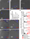
Interaction between osteocytes and S. aureus ex vivo and in clinical PJI specimens. (A and B) Presence of S. aureus (white arrowheads) in osteocyte (Ot)-occupied lacunae in bone tissue after ex vivo infection; (C) presence of S. aureus (white arrowhead) in empty lacunae (EL); (D) isotype-matched control immunostaining of infected bone tissue; (E) upregulation of mRNA levels of CXCL1, CXCL8, CXCL9, CXCL10, and CXCL11 in infected bone samples in comparison to control samples (the y axis represents log2 values of mRNA fold change [FC] and mean normalized expression ± SEM from 3 independent biological replicates); (F) immunostaining targeting S. aureus (red fluorescence) in the acetabular bone of a PJI patient, with the box marked by a broken red line indicating the enlarged area in panel F′; (F′) high-magnification image of S. aureus staining of osteocytes/osteocyte lacunae (white arrowheads); (G) isotype-matched control immunostaining of PJI patient acetabular bone; (G′) high-magnification image of control staining of osteocytes/osteocyte lacunae; (H) boxplots (median with interquartile range) showing relative mRNA levels (normalized to that of the glyceraldehyde-3-phosphate dehydrogenase [GAPDH] gene) of CCL5 (P = 0.026), CXCL9 (P = 0.038), CXCL10 (P = 0.054), and CXCL11 (P = 0.0070) in bone specimens from the iliac wing and acetabulum of the PJI cohort (n = 23) and femoral fracture cohort (n = 13). P values indicate the comparisons between acetabulum bone and iliac wing bone from PJI patients. BM, bone marrow.

References

    1. Dale H, Hallan G, Espehaug B, Havelin LI, Engesaeter LB. 2009. Increasing risk of revision due to deep infection after hip arthroplasty. Acta Orthop 80:639–645. doi:10.3109/17453670903506658. - DOI - PMC - PubMed
    1. Kurtz SM, Ong KL, Lau E, Bozic KJ, Berry D, Parvizi J. 2010. Prosthetic joint infection risk after TKA in the Medicare population. Clin Orthop Relat Res 468:52–56. doi:10.1007/s11999-009-1013-5. - DOI - PMC - PubMed
    1. Shahi A, Tan TL, Chen AF, Maltenfort MG, Parvizi J. 2017. In-hospital mortality in patients with periprosthetic joint infection. J Arthroplasty 32:948–952.e1. doi:10.1016/j.arth.2016.09.027. - DOI - PubMed
    1. Zmistowski B, Karam JA, Durinka JB, Casper DS, Parvizi J. 2013. Periprosthetic joint infection increases the risk of one-year mortality. J Bone Joint Surg Am 95:2177–2184. doi:10.2106/JBJS.L.00789. - DOI - PubMed
    1. Wimmer MD, Friedrich MJ, Randau TM, Ploeger MM, Schmolders J, Strauss AA, Hischebeth GT, Pennekamp PH, Vavken P, Gravius S. 2016. Polymicrobial infections reduce the cure rate in prosthetic joint infections: outcome analysis with two-stage exchange and follow-up ≥two years. Int Orthop 40:1367–1373. doi:10.1007/s00264-015-2871-y. - DOI - PubMed

Publication types

MeSH terms