. 2018 Apr 24;9(1):1443.
doi: 10.1038/s41467-018-03961-y.

Non-invasive detection of human cardiomyocyte death using methylation patterns of circulating DNA

Affiliations

Non-invasive detection of human cardiomyocyte death using methylation patterns of circulating DNA

Hai Zemmour et al. Nat Commun. .

Abstract

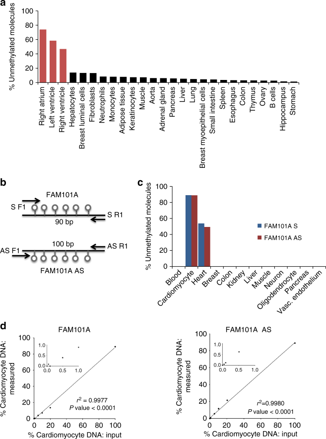
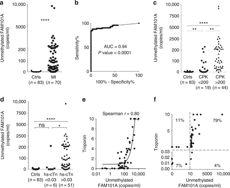
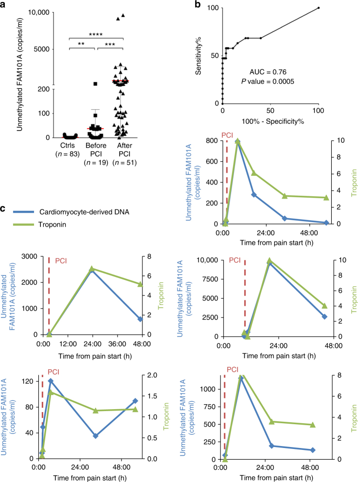
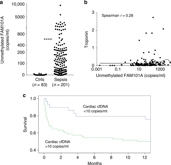
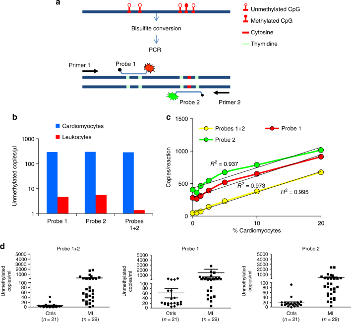
Detection of cardiomyocyte death is crucial for the diagnosis and treatment of heart disease. Here we use comparative methylome analysis to identify genomic loci that are unmethylated specifically in cardiomyocytes, and develop these as biomarkers to quantify cardiomyocyte DNA in circulating cell-free DNA (cfDNA) derived from dying cells. Plasma of healthy individuals contains essentially no cardiomyocyte cfDNA, consistent with minimal cardiac turnover. Patients with acute ST-elevation myocardial infarction show a robust cardiac cfDNA signal that correlates with levels of troponin and creatine phosphokinase (CPK), including the expected elevation-decay dynamics following coronary angioplasty. Patients with sepsis have high cardiac cfDNA concentrations that strongly predict mortality, suggesting a major role of cardiomyocyte death in mortality from sepsis. A cfDNA biomarker for cardiomyocyte death may find utility in diagnosis and monitoring of cardiac pathologies and in the study of normal human cardiac physiology and development.


Conflict of interest statement

Ju.M., R.S., B.G. and Y.D. are inventors on a patent entitled “A Novel approach for improving the detection of tissue-specific DNA: Sense and Antisense strands of bisulfite treated DNA” (patent number 62/531,990), and a patent entitled “A dual-probe digital droplet PCR strategy for specific detection of tissue-specific circulating DNA molecules” (patent number 62/531,983); H.Z., Ju.M., Jo.M., D.N., R.S., B.G. and Y.D. are inventors on a patent entitled “New set of probes for determining tissue of origin by methylation pattern” (patent number 62/531,988). The remaining authors declare no competing interests.

Figures

Fig. 1
Fig. 1
Identification of cardiomyocyte-specific DNA methylation markers. a Unmethylation levels of FAM101A locus in 27 human tissues, including left ventricle, right ventricle and right atrium (red). Data representing methylation status of all CpG sites in the locus was extracted from the Roadmap Epigenomics Consortium browser. b Structure of the FAM101A locus, used as two independent markers: FAM101A and FAM101A AS. Lollipops represent CpG sites; arrows mark positions of PCR primers; S sense marker, AS antisense marker. c Demethylated FAM101A and FAM101A AS in DNA from multiple tissues and from isolated cardiomyocytes (purchased from ScienCell Research Laboratories, San Diego, CA). See online methods for an explanation why targeted PCR yields a lower background in non-cardiac tissues compared with the Roadmap browser in panel a. d Spike in experiments for FAM101A (left) and FAM101A AS (right). Human cardiomyocyte DNA was mixed with human leukocyte DNA in the indicated proportions (0–100%), and the percentage of fully unmethylated FAM101A molecules (in which all CpG sites were converted by bisulfite) was determined
Fig. 2
Fig. 2
Cardiac cfDNA in healthy subjects and in patients with myocardial infarction. a Cardiac cfDNA (copies of fully unmethylated FAM101A /ml plasma) in samples from healthy controls (Ctrls, n = 83) and patients during STEMI (MI, n = 74 samples from 31 patients). Mann–Whitney test for controls vs. patients, p-value < 0.0001. b Receiver operating characteristic (ROC) curve for the diagnosis of STEMI by demethylated FAM101A in plasma of healthy controls and patients with MI. Area under the curve (AUC) 0.94 (95% CI = 0.9044 to 0.983), p-value < 0.0001. c Comparison of unmethylated FAM101A levels (copies/ml) in healthy controls, STEMI samples with normal Creatine Kinase (CPK < 200) and STEMI samples with high CPK (CK > 200). Kruskal-Wallis test p-value < 0.0001. Dunn’s multiple comparisons test: Controls (Ctrls) vs. STEMI with normal CPK, p-value < 0.001; Ctrls vs. STEMI with high CPK, p-value < 0.0007; STEMI with normal CPK vs. STEMI with high CPK, p-value = 0.0068. d Comparison of unmethylated FAM101A levels in healthy controls, samples from STEMI patients with normal levels of high-sensitive cardiac troponin T (hs-cTn) (<0.03), and samples from STEMI patients with high levels of hs-cTn (>0.03). Kruskal–Wallis test p-value < 0.0001. Dunn’s multiple comparisons test: Ctrls vs. STEMI with normal hs-cTn, p-value = 0.6863; Ctrls vs. STEMI with high hs-cTn, p-value < 0.0001; STEMI with normal hs-cTn vs. STEMI with high hs-cTn (>0.03), p-value = 0.0307. e Spearman correlation between cardiac cfDNA and troponin levels in 57 STEMI patients. Curved line, non linear (quadratic) fit. f XY Scatter plot for cardiac cfDNA levels vs. cardiac troponin. Quadrants indicate negative and positive hs-cTn, and negative and positive cardiac cfDNA. Numbers indicate the percentage of samples in each quadrant
Fig. 3
Fig. 3
Cardiac cfDNA dynamics during STEMI and after angioplasty. a Comparison of unmethylated FAM101A levels in healthy controls, and STEMI samples before and after PCI. Kruskal–Wallis test p-value < 0.0001. Dunn’s multiple comparisons test: Ctrls vs. pre-PCI, p-value = 0.0082; Ctrls vs. post-PCI (>0.03), p-value < 0.0001; per- vs post-PCI, p-value = 0. 0005. Horizontal red and gray lines represent the average value and standard deviation of cardiac cfDNA among the samples from each group. b ROC curve for the diagnosis of STEMI by cardiac cfDNA in healthy individuals versus STEMI patients prior to intervention. Area under the curve (AUC) 0.76, p-value = 0.0005. c Time course of cardiac cfDNA and troponin levels in representative individual patients. Vertical dashed lines indicate PCI time
Fig. 4
Fig. 4
Cardiac cfDNA in sepsis. a Levels of cardiac cfDNA in healthy controls (n = 83) and patients with sepsis (n = 201). Mann–Whitney test for controls vs. patients, p-value < 0.0001. b Lack of correlation between cardiac cfDNA and troponin. Curved line represents non-linear (quadratic) fit. Spearman r coefficient of correlation was calculated. c Kaplan–Meier plot showing correlation of cardiac cfDNA to patient survival
Fig. 5
Fig. 5
Detection of cardiac cfDNA using digital droplet PCR. a Schematic of approach for ddPCR-based detection of methylation status of multiple adjacent cytosines. A signal from two probes in the same droplet reflects lack of methylation in five adjacent cytosines in the same original DNA strand. b Signal from cardiomyocyte and leukocyte DNA based on individual or dual probes. Scoring only dual probe signals drastically reduces noise from leukocyte DNA. c Spike-in experiment assessing sensitivity and linearity of signal from cardiomyocyte DNA diluted in leukocyte DNA. The use of dual probe enhances linearity and reduces baseline signal. x axis shows both the % of cardiac DNA diluted into blood DNA, and the absolute number of cardiomyocte genomes present in each sample. d Measurement of cardiac cfDNA in plasma of healthy adult and patients with myocardial infarction. The use of dual probes reduces the baseline signal in healthy plasma. Horizontal lines represent average and standard deviation of cfDNA values among the samples in each group

References

    1. Hickman PE, et al. Cardiac troponin may be released by ischemia alone, without necrosis. Clin. Chim. Acta. 2010;411:318–323. doi: 10.1016/j.cca.2009.12.009. - DOI - PubMed
    1. Michielsen EC, Wodzig WK, Van Dieijen-Visser MP. Cardiac troponin T release after prolonged strenuous exercise. Sports Med. 2008;38:425–435. doi: 10.2165/00007256-200838050-00005. - DOI - PubMed
    1. Roca E, et al. The Dynamics of Cardiovascular Biomarkers in non-Elite Marathon Runners. J. Cardiovasc Transl. Res. 2017;10:206–208. doi: 10.1007/s12265-017-9744-2. - DOI - PubMed
    1. Landesberg G, Jaffe AS. ‘Paradox’ of troponin elevations after non-cardiac surgery. Br. J. Anaesth. 2015;114:863–865. doi: 10.1093/bja/aev068. - DOI - PubMed
    1. Alcalai R, et al. Acute coronary syndrome vs nonspecific troponin elevation: clinical predictors and survival analysis. Arch. Intern. Med. 2007;167:276–281. doi: 10.1001/archinte.167.3.276. - DOI - PubMed

Publication types

MeSH terms