. 2018 Apr 24;9(1):1629.
doi: 10.1038/s41467-018-04107-w.

Feed-forward alpha particle radiotherapy ablates androgen receptor-addicted prostate cancer

Affiliations

Feed-forward alpha particle radiotherapy ablates androgen receptor-addicted prostate cancer

Michael R McDevitt et al. Nat Commun. .

Abstract

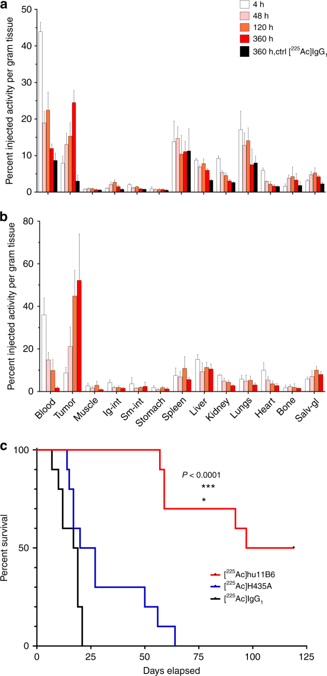
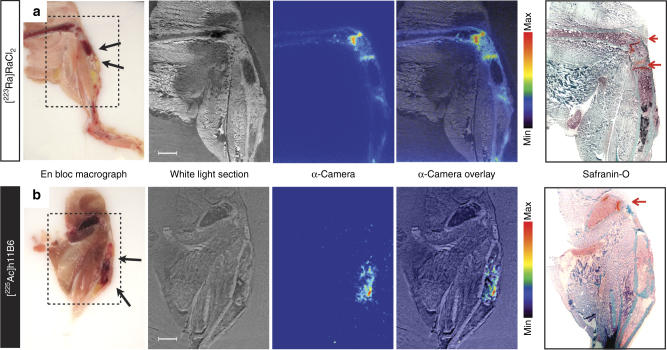
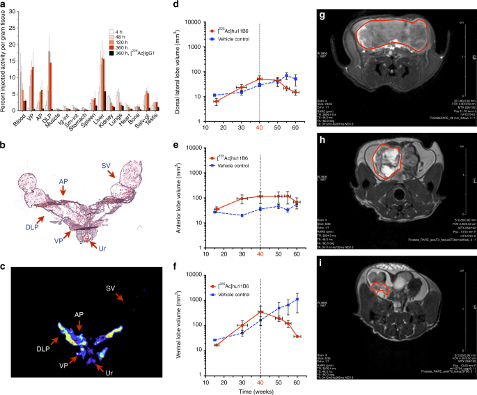
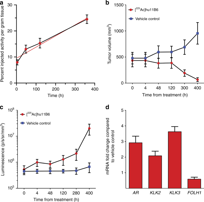
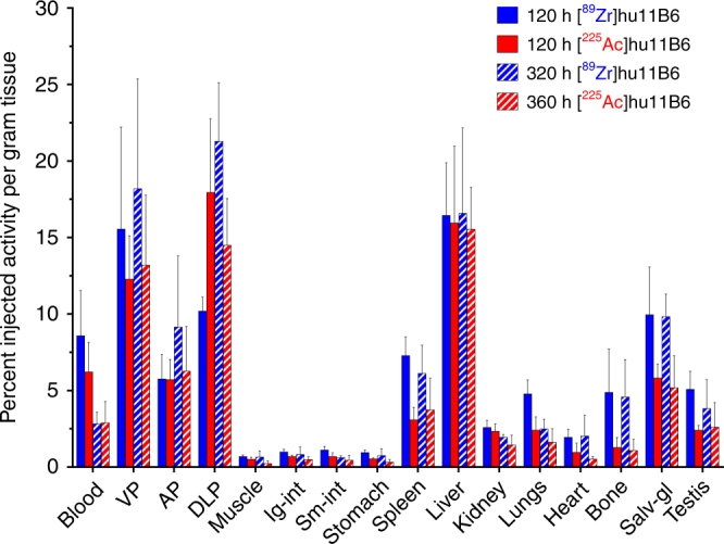
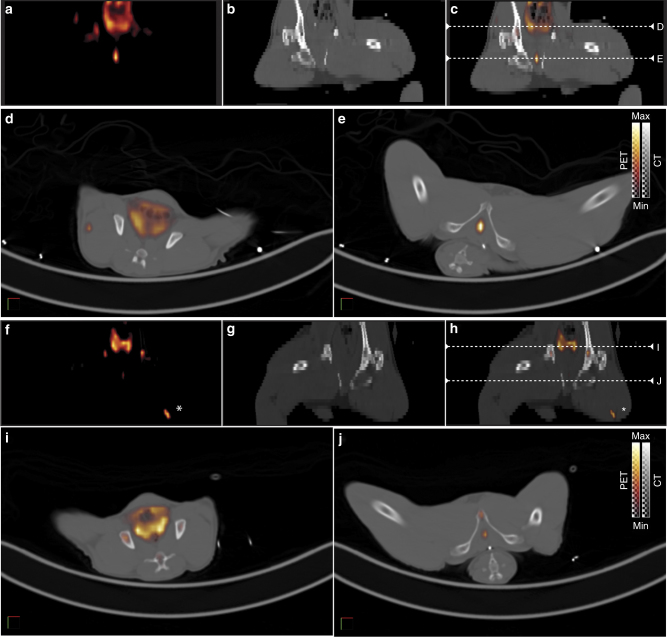
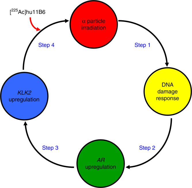
Human kallikrein peptidase 2 (hK2) is a prostate specific enzyme whose expression is governed by the androgen receptor (AR). AR is the central oncogenic driver of prostate cancer (PCa) and is also a key regulator of DNA repair in cancer. We report an innovative therapeutic strategy that exploits the hormone-DNA repair circuit to enable molecularly-specific alpha particle irradiation of PCa. Alpha-particle irradiation of PCa is prompted by molecularly specific-targeting and internalization of the humanized monoclonal antibody hu11B6 targeting hK2 and further accelerated by inherent DNA-repair that up-regulate hK2 (KLK2) expression in vivo. hu11B6 demonstrates exquisite targeting specificity for KLK2. A single administration of actinium-225 labeled hu11B6 eradicates disease and significantly prolongs survival in animal models. DNA damage arising from alpha particle irradiation induces AR and subsequently KLK2, generating a unique feed-forward mechanism, which increases binding of hu11B6. Imaging data in nonhuman primates support the possibility of utilizing hu11B6 in man.


Conflict of interest statement

Authors D.L.J.T., S.E.S., H.L. and D.U. are shareholders of Diaprost Inc., which owns the antibody hu11B6. The remaining authors declare no competing interests.

Figures

Fig. 1
Fig. 1
Pharmacokinetics and pharmacodynamics of [225Ac]hu11B6 in xenograft models of prostate cancer. Full [225Ac]hu11B6 biodistribution data set of (a) LNCaP-AR and (b) VCaP s.c. xenograft models, where the significantly higher tumor accumulation of [225Ac]hu11B6 can be noted in the VCaP model as a result of higher target expression. c Kaplan–Meier plot comparing survival of LNCaP-AR s.c. tumor models (n = 10 per group) treated with a single 300 nCi dose of [225Ac]hu11B6 (red line), 300 nCi non-internalizing hK2 targeting antibody [225Ac]hu11B6-H435A (blue line), or 300 nCi of an alpha-particle labeled non-specific antibody [225Ac]huIgG1 (black line). These results demonstrate that effective hK2-targeted alpha particle delivery by hu11B6 depends on both antigen specificity and FcRn-mediated internalization of the immunoglobulin
Fig. 2
Fig. 2
Comparative microdistribution of [223Ra]Cl2 and [225Ac]hu11B6 in mice bearing intratibial LNCaP-AR disease showed a differential uptake in vivo. Autoradiograms of (a) [223Ra]Cl2 and (b) [225Ac]hu11B6 demonstrated distinctly different microdistributions of activity. While [225Ac]hu11B6 localized directly to the tumor (indicated by black arrows in macrographs), [223Ra]Cl2 showed accumulation in apposite bone surface surrounding the lesion, as well active sites of both bone modeling and remodeling (n.b., disease is comprised of both osteolytic and osteoblastic bone lesions). Sequential tissue sections developed using Safranin-O, which differentiates mineral bone (green colored staining effect) from proteoglycans and cartilage (red-orange colored staining effect). The mineralizing front behind the growth plate at the epiphyses (red arrow) as shown in the Safranin-O stain show intense 223Ra uptake (Fig. 2a); antibody-targeted 225Ac does not accumulate at these sites (Fig. 2b)
Fig. 3
Fig. 3
Specific and potent therapeutic effect of [225Ac]hu11B6 in the Hi-Myc x pb_KLK2 mouse model that expresses human hK2 in the prostate. a The pharmacokinetic profile of [225Ac]hu11B6 was determined in a biodistribution experiment using genetically modified mice (Hi-Myc x pb_KLK2) with inherent prostate-specific expression of human hK2 and spontaneous development of prostate adenocarcinoma. This biodistribution data set shows increasing accumulation of drug in the ventral prostate (VP), dorsal-lateral prostate (DLP), and anterior prostate (AP) lobes with time (n.b., the VP and DLP exhibit higher expressions of hK2 and showed higher [225Ac]hu11B6 uptake compared to AP tissue). H&E (b) and autoradiography (c) of a tissue section (encompassing all three prostate lobes, urethra and seminal vesicles) from a pb_KLK2 x Hi-Myc injected with [225Ac]hu11B6. Radioactivity is only associated with the prostate lobes and not with seminal vesicles (SV) and radiosensitive bystander urothelium (Ur). In a therapeutic study of [225Ac]hu11B6, disease was monitored by volumetric MRI measurements of the individual lobes of Hi-Myc x pb_KLK2 mice. Mice underwent longitudinal MRI commencing at 15–16 weeks of age and received a single 300 nCi dose of [225Ac]hu11B6 at week 40. Lobe volumes are plotted vs. time following the effect of alpha irradiation on the DLP (d), AP (e), and VP (f) respectively. Vehicle-treated animals served as control for tumor progression. The results show that one injection of [225Ac]hu11B6 significantly decreased tumor burden and inhibited disease progression in this GEM model of locally advanced disease. MR images of a representative animal confirms the significant reduction in tumor volume as shown at baseline (pre-treatment) (g) and at 4 (h) and 8 (i) weeks following a single injection of [225Ac]hu11B6
Fig. 4
Fig. 4
Alpha particles promote a feed-forward oncoaddictive effect that promulgates lethality. The increasing uptake of [225Ac]hu11B6 is shown as %IA/g (red-filled circles with a blue connecting line) in LNCaP-AR tumor at four time-points after IV administration. This escalating rate of drug accumulation in tumor is noted by the linear regression fit (solid red line) with slope = 0.037 ± 0.001 %IA/g/h. Therapeutic efficacy of a single administration of [225Ac]hu11B6 is evidenced by the decreasing tumor volumes compared with vehicle-treated controls where the trajectory of untreated tumor volumes steadily increase with time. c These data report the measured AR-driven luciferase bioluminescence signal (photons/s/mm2/sr) vs. time in the treated and control animals. Note that while the alpha irradiated tumor volume decreases (see panel 4B) with time while AR-driven BLI signal increases. These data show upregulation of AR expression as a consequence of alpha irradiation with [225Ac]hu11B6. RT-PCR analysis of AR and AR-driven genes in tumor tissues collected from [225Ac]hu11B6- and vehicle-treated mice at 400 h. The fold-change in tumor gene activity of treated animals normalized to non-treated tumors shows increased expression of AR, KLK2, and KLK3, but not FOLH1
Fig. 5
Fig. 5
[89Zr]hu11B6 is a surrogate PET reporter for [225Ac]hu11B6 distribution in vivo. A pharmacokinetic comparison of [89Zr]hu11B6 and [225Ac]hu11B6 distribution in Hi-Myc x pb_KLK2 mice shows that tissue uptake and blood clearance of the 89Zr-labeled diagnostic PET hu11B6 agent is comparable to the alpha emitting 225Ac-labeled therapeutic analog at both time points investigated. This analogous pharmacokinetic profile suggests that [89Zr]hu11B6-PET can be utilized as a reporter for [225Ac]hu11B6 therapy in vivo. The tumor accumulation of both agents in all three prostate lobes is similar
Fig. 6
Fig. 6
hK2-target specificity is imaged in nonhuman primate using [89Zr]hu11B6 PET/CT. Prostate localization of diagnostic anti-hK2 tracer, [89Zr]hu11B6, imaged in a representative healthy adult male cynomolgous monkey (9 y.o.; 9.9 kg) at 8 d (a–e) and 16 d (f–j) post IV administration. (a–c, f–h) PET, CT and fusion coronal images showing the location of two axial planes (white dashed lines) for corresponding axial plane images of the prostate (d, i) and epididymis (e, j). Nonspecific uptake at 16 d in left thigh (f, h) appears to localize to site of anesthetic administration (white asterisk)
Fig. 7
Fig. 7
The mechanism of action in alpha particle-promoted feed-forward oncoaddiction. The interaction of α-particles with tumor tissue produces DNA damage. The ensuing transcriptional response prompts the hormone-DNA circuit to upregulate AR expression. KLK2 expression subsequently upregulates due to AR and produces more hk2 target epitope for [225Ac]hu11B6

References

    1. Jurcic JG, et al. Targeted α-particle immunotherapy for myeloid leukemia. Blood. 2002;100:1233–1239. - PubMed
    1. Parker C, et al. ALSYMPCA Investigators. Alpha emitter radium-223 and survival in metastatic prostate cancer. N. Engl. J. Med. 2013;369:213–223. doi: 10.1056/NEJMoa1213755. - DOI - PubMed
    1. Jurcic J, et al. Phase I trial of alpha-particle immunotherapy with 225Ac-lintuzumab and low-dose cytarabine in patients age 60 or older with untreated acute myeloid leukemia. J. Nucl. Med. 2017;58:456.
    1. McDevitt MR, et al. Tumor therapy with targeted atomic nanogenerators. Science. 2001;294:1537–1540. doi: 10.1126/science.1064126. - DOI - PubMed
    1. Nikula TN, et al. Alpha-emitting bismuth cyclohexylbenzyl DTPA constructs of recombinant humanized anti-CD33 antibodies: pharmacokinetics, bioactivity, toxicity and chemistry. J. Nucl. Med. 1999;40:166–176. - PubMed

Publication types