. 2018 Apr 24;9(1):1640.
doi: 10.1038/s41467-018-04049-3.

Control of mechanical pain hypersensitivity in mice through ligand-targeted photoablation of TrkB-positive sensory neurons

Affiliations

Control of mechanical pain hypersensitivity in mice through ligand-targeted photoablation of TrkB-positive sensory neurons

Rahul Dhandapani et al. Nat Commun. .

Abstract

Mechanical allodynia is a major symptom of neuropathic pain whereby innocuous touch evokes severe pain. Here we identify a population of peripheral sensory neurons expressing TrkB that are both necessary and sufficient for producing pain from light touch after nerve injury in mice. Mice in which TrkB-Cre-expressing neurons are ablated are less sensitive to the lightest touch under basal conditions, and fail to develop mechanical allodynia in a model of neuropathic pain. Moreover, selective optogenetic activation of these neurons after nerve injury evokes marked nociceptive behavior. Using a phototherapeutic approach based upon BDNF, the ligand for TrkB, we perform molecule-guided laser ablation of these neurons and achieve long-term retraction of TrkB-positive neurons from the skin and pronounced reversal of mechanical allodynia across multiple types of neuropathic pain. Thus we identify the peripheral neurons which transmit pain from light touch and uncover a novel pharmacological strategy for its treatment.


Conflict of interest statement

The authors declare no competing interests.

Figures

Fig. 1
Fig. 1
TrkB-positive sensory neurons are putative mechanoreceptors. ad Double immunofluorescence of DRG sections from TrkBCreERT2::Rosa26RFP mice with a NF200, b Ret, visualized using TrkBCreERT2::Rosa26RFP::RetEGFP triple transgenic mice, c IB4, d quantification of staining on mouse DRG sections; TrkB+ cells account for ~10% of all DRG neurons and all co-express NF200 or NF200 + ReteGFP, while they are negative for IB4, CGRP, and TH. eh Double immunofluorescence of human DRG sections stained with antibodies against TrkB and e NF200, f Ret, g TrkA. h Size distribution for human DRG neurons expressing TrkB, NF200, and TrkA. i Section through the lumbar spinal cord of TrkBCreERT2::AvilmCherry mice stained with IB4. j TrkB+ lanceolate endings in a section of the back hairy skin of TrkBCreERT2::Rosa26SnapCaaX labeled with Snap Cell TMRstar (red), NF200 (green), and DAPI (blue). k Section from the glabrous skin of TrkBCreERT2::Rosa26ChR2YFP (red) stained with anti-S100 a marker for Meissner’s corpuscles (green) and DAPI (blue) showing TrkB+ innervation. l Section from human glabrous skin stained with antibodies against TrkB (red) and NF200 (green), and DAPI (blue). Scale bars, ac and i 50 μm, eg and jl 40 μm.
Fig. 2
Fig. 2
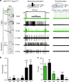
TrkB-positive sensory neurons are myelinated low-threshold mechanoreceptors. In vitro skin nerve preparation from TrkBCreERT2::Rosa26ChR2 mice showing a representative responses to 10 Hz stimulation with blue light, b the minimal force required to elicit an action potential in the indicated fiber type, and c the conduction velocities of the fiber types. Green bar represents TrkB+ afferents, n number indicated in brackets. Error bars represent SEM
Fig. 3
Fig. 3
Diphtheria toxin-mediated ablation of TrkB+ sensory neurons. Immunostaining of DRG sections of TrkBCreERT2::AviliDTR mice with an antibody against the diphtheria toxin receptor (red) from a untreated mice and b after i.p. injections of diphtheria toxin. c Quantification of DRG sections indicating a ~90% decrease in TrkBDTR and TrkBmRNA cells after ablation and 36% reduction in NF200+ neurons without affecting other subpopulations. dl Behavioral responses in littermate control mice (AviliDTR, black bars) and TrkBCreERT2::AviliDTR mice (white bars) showing no differences in responses before and after ablation in the acetone drop test (t-test; p = 0.800) on d glabrous and e hairy skin, f hot plate test (t-test; p = 0.914), g radiant heat test on glabrous and hairy skin (t-test; p = 0.263), h grip test (t-test; p = 0.484), i pin-prick test (t-test; p = 0.691), j tape test (t-test; p = 0.776), and k punctate mechanical flick test applied to the hairy back skin (t-test; p = 0.196). l Ablated mice show a reduction in sensitivities to cotton swab (t-test, p = 0.002). Scale bars in a, b 50 μm, error bars indicate SEM
Fig. 4
Fig. 4
TrkB+ neurons are necessary and sufficient to convey mechanical allodynia after nerve injury. a Schematic of CFA injection and behavior tests following ablation of TrkB+ neurons. Mechanical hypersensitivity in control AviliDTR (black bar), TrkBCreERT2::AviliDTR (white bar), and sham-injected (gray bar) mice 48 h after CFA injections as measured by b von Frey filaments (t-test, p = 0.886), c dynamic brush stimuli (t-test; p = 0.537), and d cotton swab stimuli (t-test; p = 0.242). All mice received two diphtheria toxin injections 7 days and 10 days before CFA treatment. e Paw withdrawal frequencies in control (Rosa26ChR2 mice without Cre, black bar), and CFA-injected (white bar) or sham (grey bar)-injected paw of TrkBCreERT2::Rosa26ChR2 mice upon stimulation with 473 nm blue light. No significant differences under baseline conditions and 48 h after CFA injection (Mann–Whitney test; p = 0.886). f Schematic of SNI and behavioral tests following ablation of TrkB+ neurons. g von Frey mechanical thresholds indicating that ablation of TrkB+ neurons abolished the development of mechanical allodynia after SNI in TrkBCreERT2::AviliDTR mice (white circles) as compared to AviliDTR controls (black circles) (n = 7 for both sets, two-way RM ANOVA; p = 0.001 followed by a Bonferroni post hoc test). Sham-operated mice (grey circles) did not develop mechanical hypersensitivity. h, i Reduced dynamic allodynia in ablated TrkBCreERT2::AviliDTR mice (white bar) as compared to littermate controls (black bar; t-test p = 0.016) stimulated with a h brush or i cotton swab. Sham-operated mice did not develop dynamic allodynia. j Nociceptive behavior evoked by optogenetic stimulation of the paws of Rosa26ChR2 mice without Cre (black bars) and TrkBCreERT2::Rosa26ChR2 (white bars) mice after SNI, or ipsilateral paws (grey bars) of sham-operated mice (two-way RM ANOVA; p = 0.001). k Cross sections of lumbar spinal cord from TrkBCreERT2::Rosa26ChR2 mice labeled for c-fos (red) and IB4 (green) after 1 min exposure to 15 Hz blue light. Representation of section taken from an uninjured mouse and a section from an injured mouse at 7 days post SNI. l Quantification of the number of c-fos positive cells in laminae I, II, and III/V of the lumbar spinal cord within a 40 μm section. Data are shown for SNI, non-injured and sham-operated TrkBCreERT2::Rosa26ChR2 mice with or without light, and control SNI Rosa26ChR2 mice without Cre. Baseline indicates pre-ablation and pre-treatment. Error bars indicate SEM. Scale bars in k, 40 μm
Fig. 5
Fig. 5
Optopharmacological targeting of TrkB+ neurons with BDNFSNAP. a Labeling (inset) and quantification of dissociated DRG from TrkBCreERT2::Rosa26RFP mice with BDNFSNAP shows substantial overlap of BDNFSNAP binding to TrkB+ cells (n = 4, 450 cells). b Schematic representation of BDNFSNAP-IR700 injection and photoablation. c BDNFSNAP-IR700-mediated photoablation of the paw of SNI mice results in a dose-dependent reversal of mechanical hypersensitivity as assayed with von Frey filaments (n = 10, two-way RM ANOVA; p = 0.003 followed by a Bonferroni post hoc test) and d dynamic brush stimuli (t-test; p = 0.016). e Hypersensitivity to cotton swab is also reversed by photoablation (t-test: p = 0.042). f BDNFSNAP-IR700-mediated photoablation reverses mechanical allodynia in the streptozotocin (STZ) model of diabetic neuropathy (n = 5, two-way RM ANOVA; p = 0.007 followed by a Bonferroni post hoc test. Open circles; 5 µM BDNFSNAP-IR700 at 200 J/cm2, closed circles, 5 µM IR700 at 200 J/cm2). g BDNFSNAP-IR700-mediated photoablation reverses mechanical allodynia in the paclitaxel (PTX) model of chemotherapy-induced neuropathy (n = 5, two-way RM ANOVA; p = 0.027 followed by a Bonferroni post hoc test. Open circles; 5 µM BDNFSNAP-IR700 at 200 J/cm2, closed circles, 5 µM IR700 at 200 J/cm2). Error bars indicate SEM
Fig. 6
Fig. 6
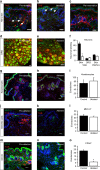
BDNFSNAP-IR700 photoablation promotes local retraction of TrkB+ afferents. ac Substantial loss of TrkBCreERT2 positive afferents (red), but persistence of other fibers (green) upon BDNFSNAP-IR700-mediated photoablation. a Innervation of paw hairy skin prior to ablation, arrows show lanceolate endings. b Loss of TrkBCreERT2 afferents after ablation, arrows show PGP9.5 fibers. c Re-innervation of skin by TrkBCreERT2 afferents at 24 days post ablation. d DRG section from control TrkBCreERT2 mouse labeled for RFP (red) and NF200 (green). e DRG section from photoablated TrkBCreERT2 mouse labeled for RFP (red) and NF200 (green). f Quantification of the proportion of hair follicle innervation and DRG neurons positive for TrkB following photoablation in the paw and the quantification of PGP9.5+ free nerve endings showing the numbers of free nerves remain unaffected. Representative skin sections from control and BDNFSNAP-IR700 photoablated mice labeled with the indicated antibodies. TrkB-positive cells are indicated in red and DAPI-positive nuclei in blue. g, h Keratinocytes labeled with K14 (green). i Quantification of the number of K14+ cells. j, k Dendritic cells and dermal antigen-presenting cells labeled with MHC-II (green) and l the quantification for MHC-II+ cells. m, n Mast cells and epithelial and endothelial progenitor cells labeled with CD34 (green). o Quantification of the number of K14+ cells. Scale bars, 40 µm. Error bars indicate SEM
Fig. 7
Fig. 7
Photoablation of the sciatic nerve shows requirement of TrkB+ neurons in mediating allodynia. af Representative images of the sciatic nerve labeled for TrkB in green, DAPI in blue, and CD34 or S100 in red. Longitudinal sections of the nerve show reduction in TrkB+ fibers, but no detectable CD34 in a non-ablated control and b photoablated mice. Cross-section of the sciatic nerve from c control and d photoablated mice shows reduction in TrkB+ fibers. No co-localization between TrkB+ fibers and S100+ cells in e longitudinal sections and f cross-section of the nerve. g Quantification of the numbers of TrkB+ fibers and DAPI labeled cells in cross sections of sciatic nerve. Behavioral sensitivity following BDNFSNAP-IR700-mediated ablation in the sciatic nerve: h acetone drop test (t-test; p = 0.151), i radiant heat test (t-test; p = 0.829), and j pin-prick test (t-test; p = 0.548) are not altered by nerve photoablation. However, sensitivity to k cotton swab (t-test; p = 0.001) in control animals, and l light-evoked behavior in TrkBCreERT2::Rosa26ChR2 mice with SNI, are reduced by nerve photoablation (two-way RM ANOVA; p = 0.002). White bars 5 µM BDNFSNAP-IR700 at 200 J/cm2, black bars 5 µM IR700 at 200 J/cm2. Baseline indicates pre-ablation and pre-treatment. Error bars indicate SEM. Scale bars, ad 40 μm, e 10 μm

Similar articles

Cited by

References

    1. Treede RD, Meyer RA, Raja SN, Campbell JN. Peripheral and central mechanisms of cutaneous hyperalgesia. Prog. Neurobiol. 1992;38:397–421. doi: 10.1016/0301-0082(92)90027-C. - DOI - PubMed
    1. Koltzenburg M. Neural mechanisms of cutaneous nociceptive pain. Clin. J. Pain. 2000;16:S131–S138. doi: 10.1097/00002508-200009001-00004. - DOI - PubMed
    1. Duan B, et al. Identification of spinal circuits transmitting and gating mechanical pain. Cell. 2014;159:1417–1432. doi: 10.1016/j.cell.2014.11.003. - DOI - PMC - PubMed
    1. Peirs C, et al. Dorsal horn circuits for persistent mechanical pain. Neuron. 2015;87:797–812. doi: 10.1016/j.neuron.2015.07.029. - DOI - PMC - PubMed
    1. Foster E, et al. Targeted ablation, silencing, and activation establish glycinergic dorsal horn neurons as key components of a spinal gate for pain and itch. Neuron. 2015;85:1289–1304. doi: 10.1016/j.neuron.2015.02.028. - DOI - PMC - PubMed

Publication types

MeSH terms