. 2018 Jun;83(6):1105-1124.
doi: 10.1002/ana.25241.

Congenital Titinopathy: Comprehensive characterization and pathogenic insights

Emily C Oates  1   2   3   4 Kristi J Jones  2   3 Sandra Donkervoort  5 Amanda Charlton  3   6 Susan Brammah  7 John E Smith 3rd  8 James S Ware  9   10 Kyle S Yau  11 Lindsay C Swanson  12 Nicola Whiffin  9   10 Anthony J Peduto  13   14 Adam Bournazos  2   3 Leigh B Waddell  2   3 Michelle A Farrar  15   16 Hugo A Sampaio  15   16 Hooi Ling Teoh  15   16 Phillipa J Lamont  17 David Mowat  16   18 Robin B Fitzsimons  19 Alastair J Corbett  20 Monique M Ryan  21   22   23 Gina L O'Grady  2   3   24 Sarah A Sandaradura  2   3 Roula Ghaoui  2   3 Himanshu Joshi  2 Jamie L Marshall  25   26 Melinda A Nolan  24 Simranpreet Kaur  2 Jaya Punetha  27   28 Ana Töpf  29 Elizabeth Harris  29 Madhura Bakshi  30 Casie A Genetti  12 Minttu Marttila  12 Ulla Werlauff  31 Nathalie Streichenberger  32   33 Alan Pestronk  34   35 Ingrid Mazanti  36 Jason R Pinner  37 Carole Vuillerot  38   39 Carla Grosmann  40 Ana Camacho  41 Payam Mohassel  5 Meganne E Leach  5 A Reghan Foley  5 Diana Bharucha-Goebel  5   42 James Collins  43 Anne M Connolly  44 Heather R Gilbreath  45 Susan T Iannaccone  46   47 Diana Castro  46   47 Beryl B Cummings  25   48   49 Richard I Webster  50 Leïla Lazaro  51 John Vissing  52 Sandra Coppens  53   54 Nicolas Deconinck  54 Ho-Ming Luk  55 Neil H Thomas  56 Nicola C Foulds  57 Marjorie A Illingworth  56 Sian Ellard  58   59 Catriona A McLean  60   61 Rahul Phadke  1   62 Gianina Ravenscroft  63 Nanna Witting  64 Peter Hackman  65 Isabelle Richard  66 Sandra T Cooper  2   3 Erik-Jan Kamsteeg  67 Eric P Hoffman  27   28 Kate Bushby  29 Volker Straub  29 Bjarne Udd  65   68   69   70 Ana Ferreiro  71   72 Kathryn N North  2   22 Nigel F Clarke  2   3 Monkol Lek  25   48 Alan H Beggs  12 Carsten G Bönnemann  5 Daniel G MacArthur  25   48 Henk Granzier  8 Mark R Davis  73 Nigel G Laing  63
Affiliations

Congenital Titinopathy: Comprehensive characterization and pathogenic insights

Emily C Oates et al. Ann Neurol. 2018 Jun.

Abstract

Objective: Comprehensive clinical characterization of congenital titinopathy to facilitate diagnosis and management of this important emerging disorder.

Methods: Using massively parallel sequencing we identified 30 patients from 27 families with 2 pathogenic nonsense, frameshift and/or splice site TTN mutations in trans. We then undertook a detailed analysis of the clinical, histopathological and imaging features of these patients.

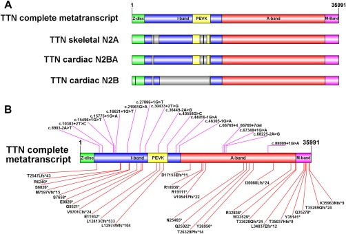
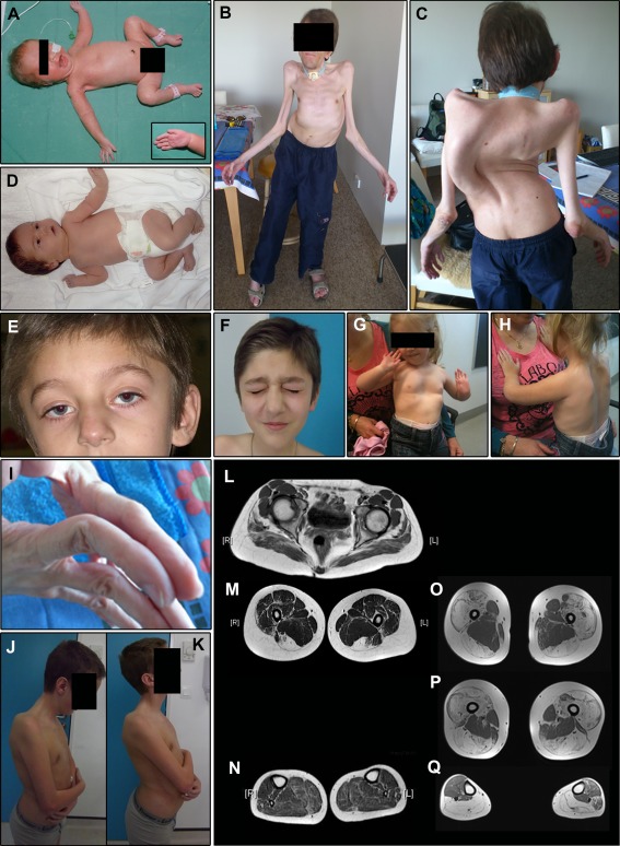
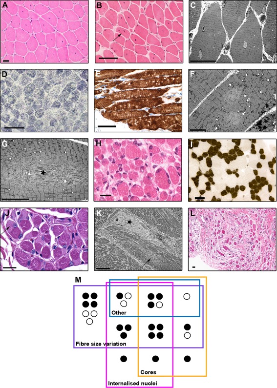
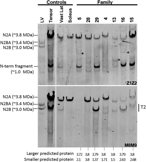
Results: All patients had prenatal or early onset hypotonia and/or congenital contractures. None had ophthalmoplegia. Scoliosis and respiratory insufficiency typically developed early and progressed rapidly, whereas limb weakness was often slowly progressive, and usually did not prevent independent walking. Cardiac involvement was present in 46% of patients. Relatives of 2 patients had dilated cardiomyopathy. Creatine kinase levels were normal to moderately elevated. Increased fiber size variation, internalized nuclei and cores were common histopathological abnormalities. Cap-like regions, whorled or ring fibers, and mitochondrial accumulations were also observed. Muscle magnetic resonance imaging showed gluteal, hamstring and calf muscle involvement. Western blot analysis showed a near-normal sized titin protein in all samples. The presence of 2 mutations predicted to impact both N2BA and N2B cardiac isoforms appeared to be associated with greatest risk of cardiac involvement. One-third of patients had 1 mutation predicted to impact exons present in fetal skeletal muscle, but not included within the mature skeletal muscle isoform transcript. This strongly suggests developmental isoforms are involved in the pathogenesis of this congenital/early onset disorder.

Interpretation: This detailed clinical reference dataset will greatly facilitate diagnostic confirmation and management of patients, and has provided important insights into disease pathogenesis. Ann Neurol 2018;83:1105-1124.


Conflict of interest statement

Nothing to report.

Figures

Figure 1
Figure 1
Schematic representation of titin isoforms and location of patient mutations. (A) Size (relative to number of amino acids) of the 4 main titin regions encoded by the inferred complete metatranscript (Refseq transcript NM_001267550.1), the single characterized skeletal muscle isoform N2A (Refseq transcript NM_133378.4), the principal cardiac long isoform N2BA (NM_001256850.1) and the principle cardiac short isoform N2B (NM_003319.4). The Z‐disc region of titin (green) interacts with α‐actinin, telethonin, and other Z‐disc–related proteins. The I‐band region (blue) contains multiple tandem immunoglobulin‐like domains and the “PEVK” domain (yellow), which is rich in proline (P), glutamic acid (E), valine (V), and lysine (K). The PEVK domain unravels when stretched, giving titin its elastic properties. The A‐band region (red) contains multiple myosin and C‐protein binding sites, and alternating fibronectin type III and immunoglobulin repeats that form a shape that complements myosin. The M‐band region (purple) is encoded by the last 6 exons [M‐band exon (Mex) exons 1–6 (exons 359–364)] and contains a kinase domain, immunoglobulin domains, and binding sequences for calpain 3, obscurin, MURF‐1, and numerous other proteins, along with additional unique sequences. The gray regions shown within N2A, N2BA, and N2B are those included in the metatranscript but not present within the relevant isoform. Thick black lines within the gray regions represent smaller subregions that are retained by the isoform, but are not large enough to show up as a colored segment. (B) Location of each of the mutations identified in our clinical analysis cohort (Families 1–27) mapped to the inferred complete metatranscript. Splice site mutations are shown above the transcript image. Frameshift and nonsense mutations are shown below the transcript. Supplementary Table 1 shows which mutations are included in N2A, N2BA and/or N2B. (Schematic images were created using Illustrator for Biological Sequences.)
Figure 2
Figure 2
Examples of common and clinically significant features. Lower limb magnetic resonance imaging (MRI) is also shown. (A) Female Family 25 proband. The pregnancy for this infant was complicated by reduced fetal movements and prenatal ultrasound detection of limb contractures. The image shows typical “frog leg” positioning due to marked congenital hypotonia and weakness. Note also the nasogastric feeding tube and reduced palmar creases (inset). This infant also had mild pulmonary stenosis and needed intubation and ventilation for a short period following delivery. (B, C) The older of the 2 affected brothers (Sibling 1) from Family 1 at age 34 years. Both brothers had congenital scoliosis, which progressed during childhood. Spinal surgery was not possible in the pictured brother due to concurrent development of severe respiratory insufficiency. His respiratory deficit was diagnosed at age 13 years following an out of hospital respiratory arrest, and he has since relied on nocturnal ventilation via a tracheostomy tube. This patient has slowly progressive mild to moderate limb weakness but remains ambulant despite significant axial involvement. He also has left ventricular cardiac dysfunction. In addition, these images demonstrate bilateral non‐congenital elbow contractures and marked generalized muscle hypotrophy. (D) Family 24 female proband who was born following a pregnancy complicated by reduced fetal movements and breech presentation. She was significantly hypotonic at birth and had congenital bilateral wrist contractures and fixed foot deformities. (E, F) Ptosis and facial weakness in the younger sibling (Sibling 2) from Family 26 during early childhood (E) and at age 12 years (F). (G, H) The same child as shown in (A) at age 2 years with a pectus excavatum chest wall deformity (G) and scapular winging (H). (I) Distal joint hypermobility in Sibling 1 from Family 1. (J, K) Reduced range of neck movement in Sibling 1 from Family 26. (L–N) Lower limb MRI result for Sibling 1 from Family 26 at age 15 years. This shows fatty infiltration of the gluteal muscles (L), severe fatty replacement of both visible hamstring muscles (complete fatty replacement of semitendinosus and incomplete but marked replacement of biceps femoris; M), relative sparing of the adductor compartment, and mild involvement of the calf and peroneal muscles (N). (O–Q) Lower limb MRI result for the Family 31 proband at age 29 years. This is one of the 4 segregation‐inconclusive cases described in this paper, but not included within the clinical analysis cohort. The features in this individual's lower limb MRI may represent the more severe end of the spectrum of muscle involvement associated with this disorder. The images show severe fatty replacement of the quadriceps and hamstrings, sparing of the adductors (also seen in the first MRI case), and severe replacement of both calves and the right peroneus, with mild fatty marbling in the anterior and lateral compartment on the left. Consent was obtained from patients/parents/legal guardians for use of the clinical photographs shown in this figure, including the non‐obscured facial photographs shown in D–F.
Figure 3
Figure 3
Examples of histopathological and ultrastructural features. All brightfield scale bars = 50 µm unless otherwise stated. (A) Centralized nuclei in the pattern of centronuclear myopathy in a hematoxylin and eosin (H&E)‐stained quadriceps section from the Family 3 female proband at age 5 years. Scale bar = 20 µm. (B) Both internalized and centralized nuclei in an H&E‐stained quadriceps section from the male proband from Family 5, at age 14 years. One fiber (arrow) has multiple internalized nuclei, a feature that was much more prominent in biopsies from older patients. This image also shows a very mild increase in endomysial connective tissue. (C) Electron micrograph (EM) image showing internalized nuclei and scattered, small, poorly defined areas of sarcomeric disruption consistent with minicores (Sibling 1/Family 1, quadriceps, age = 14 years, scale bar = 20 µm). (D) Lucent areas compatible with multiminicores in a succinate dehydrogenase–stained section from the same biopsy as shown in C. The multiminicores in D were confirmed ultrastructurally, as shown in F, which presents 2 minicores in a longitudinally orientated fiber (scale bar = 5 µm). (E) Multiminicores as discrete nonstaining foci in a longitudinally orientated paraffin section stained immunohistochemically for desmin (Sibling 2/Family 1, quadriceps, age = 10 years). (G) EM image of a myofiber containing a large centrally placed unstructured core with prominent Z‐band streaming (star; Sibling 1/Family 1, quadriceps, age = 14 years, scale bar = 10 µm). (H) Significant fiber size variation in the antemortem quadriceps biopsy taken on day 1, from the male Family 6 proband who died later the same day (scale bar = 20 µm). (I) Classical features of congenital fiber type disproportion, including T1 fibers which are > 25% smaller than T2 fibers, and T1 predominance, in the absence of other abnormalities (adenosine triphosphatase pH 4.3 section from Family 2 proband, site unknown, age = 3 years). (J) Multiple cap‐like regions (one shown with arrow) in a periodic acid Schiff–stained paraffin section (Sibling 2/Family 1, quadriceps, age = 10 years, scale bar = 20 µm). (K) EM image of a sharply demarcated cap‐ike region (star) characterized by marked myofibrillar disruption, loss of thick filaments, and thickened Z‐discs. The fiber also contains a minicore (arrow) and numerous peripheral mitochondria (Sibling 2/Family 1, quadriceps, age = 10 years, scale bar = 10 µm). (L) An area of fibrosis from patient shown in C (H&E, scale bar = 20 µm). (M) Schematic representation of the overlap between the main histopathological patterns seen in patient biopsies. “Other” refers to rarer structural abnormalities: cap‐like regions, ring, coiled, and whorled fibers, and central and peripheral mitochondrial accumulations. Black circles indicate patients with two N2A mutations. White circles indicate patients with 1 N2A mutation and 1 metatranscript‐only mutation.
Figure 4
Figure 4
Western blot analysis of patient muscle samples. Antititin antibodies were used that were specific for the titin N‐terminus (Z1Z2; top panel) or C‐terminus (M8M9; bottom panel). The 4 left lanes are controls, and the 7 right lanes are biopsies from patients (from Families 4, 5, 13, 15, and 16), or segregation‐inconclusive cases (Families 28 and 29). All patients have mutations in both of their TTN alleles that produce proteins that are predicted to vary in size. The segregation‐inconclusive cases also have 2 TTN mutations that are predicted to produce proteins of different sizes if their mutations are (as suspected) in trans. The bottom of the figure shows the expected protein mass, assuming that the wild‐type full‐length titin is 3.8MDa and that the mutant protein is reduced by the size of the missing exons(s). The largest predicted proteins are in patient biopsies from Families 28, 29, 4, 13, and 16 at nearly full size (in‐frame deletion of a single small exon near the middle of titin), consistent with their observed expression of a large titin that reacts with both Z1Z2 and M8M9. The largest predicted proteins in the remaining 2 patients (from Families 5 and 15) are slightly smaller than full‐length titin. The patient from Family 5 has a TTN truncating mutation in exon 359 reducing its size by 112KDa and eliminating the M8M9 binding site, consistent with the finding that titin in muscle from this patient reacts with Z1Z2 but not M8M9. The patient from Family 15 has a frameshift mutation in exon 364. Although this leaves the binding site for M8M9 intact, Patient 15 titin reacts only weakly with M8M9 (in contrast with the strong Z1Z2 reactivity), suggesting that the antigen availability is reduced, or perhaps that the mutant titin is degraded near its C‐terminus. The second mutant allele is, in all of the patients/cases, predicted to produce a protein that is between 1.5 and 2.7MDa (see bottom of figure) that reacts with Z1Z2 but not M8M9. Although weak bands with the expected reactivity are present in some of the patients, they are very minor relative to (nearly) full‐length titin, except for the band marked by an asterisk in the biopsy from Family 13. Also note that several patients have a Z1Z2‐positive, M8M9‐negative band at a molecular mass of ∼1.0MDa. However, this band is also seen in several control samples and is present at the same size in different patients with different mutations and therefore it is unlikely that this ∼1.0MDa band is mutation‐specific. LV = left ventricle; T2 = degradation product of titin that mainly consists of titin's A‐band segment; Vast Lat = Vastus lateralis.

Similar articles

Cited by

References

    1. Bang ML, Centner T, Fornoff F, et al. The complete gene sequence of titin, expression of an unusual approximately 700‐kDa titin isoform, and its interaction with obscurin identify a novel Z‐line to I‐band linking system. Circ Res 2001;89:1065–1072. - PubMed
    1. Labeit S, Kolmerer B. Titins: giant proteins in charge of muscle ultrastructure and elasticity. Science 1995;270:293–296. - PubMed
    1. Whiting A, Wardale J, Trinick J. Does titin regulate the length of muscle thick filaments? J Mol Biol 1989;205:263–268. - PubMed
    1. Tskhovrebova L, Trinick J. Titin: properties and family relationships. Nat Rev Mol Cell Biol 2003;4:679–689. - PubMed
    1. Ehler E, Gautel M. The sarcomere and sarcomerogenesis. Adv Exp Med Biol 2008;642:1–14. - PubMed

Publication types