

Intraventricular Neurocysticercosis: Experience and Long-Term Outcome from a Tertiary Referral Center in the United States
- PMID: 29692305
- PMCID: PMC6086198
- DOI: 10.4269/ajtmh.18-0085
Intraventricular Neurocysticercosis: Experience and Long-Term Outcome from a Tertiary Referral Center in the United States
Abstract
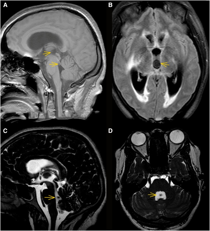
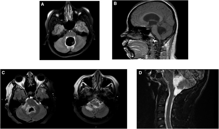
Ventricular involvement in neurocysticercosis (NCC), a common serious manifestation of NCC, has distinct clinical presentations, complications, and treatments primarily because of partial or complete obstruction of the cerebrospinal fluid (CSF) flow by Taenia solium cysts. We review the clinical course, treatments, and long-term outcomes in 23 of 121 (19.0%) total NCC patients with ventricular cysts referred to the National Institutes of Health from 1985 to the October 2017. Patients had a median age of 31.8 (range: 22.4-52.6 years), were 60.9% male, diagnosed a median of 6.5 years (range: 0.17-16 years) after immigration, and were followed for a median of 3.6 years (range: 0.1-30.5 years). Other forms and manifestations of NCC were present in 73.9% (17/23). The fourth ventricle was involved in a majority (15/23, 65.2%) resulting in hydrocephalus (73.9%), ventriculitis, and periventricular edema (7/23, 30.4%). Cystectomy was accomplished in 60.9%, usually by removal of a fourth ventricular cyst through a suboccipital craniotomy. Nonresectable cysts were treated medically. Ventriculoperitoneal shunts were inserted in 43.5% (10/23) and failed in four, three from infection. Other complications included surgically induced injuries (4/23, 17.4%) and entrapment of a lateral ventricle (2/23, 8.7%). Despite a common severe early course, 90.9% (20/22) stabilized without recurrence, 15% (3/20) complained of mild-to-moderate neurological complaints, and 15% (3/20) were significantly disabled. Four patients who underwent removal of ventricular cysts without significant other NCC and who received with no cysticidal treatment became CSF cestode antigen negative without recurrence indicating that after successful extraction of cysts, additional cysticidal treatment may not be needed.
Figures
Comment in
-
Ventricular Neurocysticercosis: A Severe Form of the Disease Waiting for Well-Designed Therapeutic Trials.Am J Trop Med Hyg. 2018 Jun;98(6):1553-1554. doi: 10.4269/ajtmh.18-0203. Epub 2018 Apr 26. Am J Trop Med Hyg. 2018. PMID: 29714161 Free PMC article. No abstract available.
References
-
- Apuzzo ML, Dobkin WR, Zee CS, Chan JC, Giannotta SL, Weiss MH, 1984. Surgical considerations in treatment of intraventricular cysticercosis. An analysis of 45 cases. J Neurosurg 60: 400–407. - PubMed
-
- Cuetter AC, Andrews RJ, 2002. Intraventricular neurocysticercosis: 18 consecutive patients and review of the literature. Neurosurg Focus 12: e5. - PubMed
-
- Madrazo I, Garcia RJ, Sandoval M, Lopez VF, 1983. Intraventricular cysticercosis. Neurosurgery 12: 148–152. - PubMed
-
- Sinha S, Sharma BS, 2012. Intraventricular neurocysticercosis: a review of current status and management issues. Br J Neurosurg 26: 305–309. - PubMed
MeSH terms
Substances
LinkOut - more resources
Full Text Sources
Other Literature Sources
Medical
