. 2018 Apr 24;2(8):941-953.
doi: 10.1182/bloodadvances.2017015008.

Red pulp macrophages in the human spleen are a distinct cell population with a unique expression of Fc-γ receptors

Affiliations

Red pulp macrophages in the human spleen are a distinct cell population with a unique expression of Fc-γ receptors

Sietse Q Nagelkerke et al. Blood Adv. .

Abstract

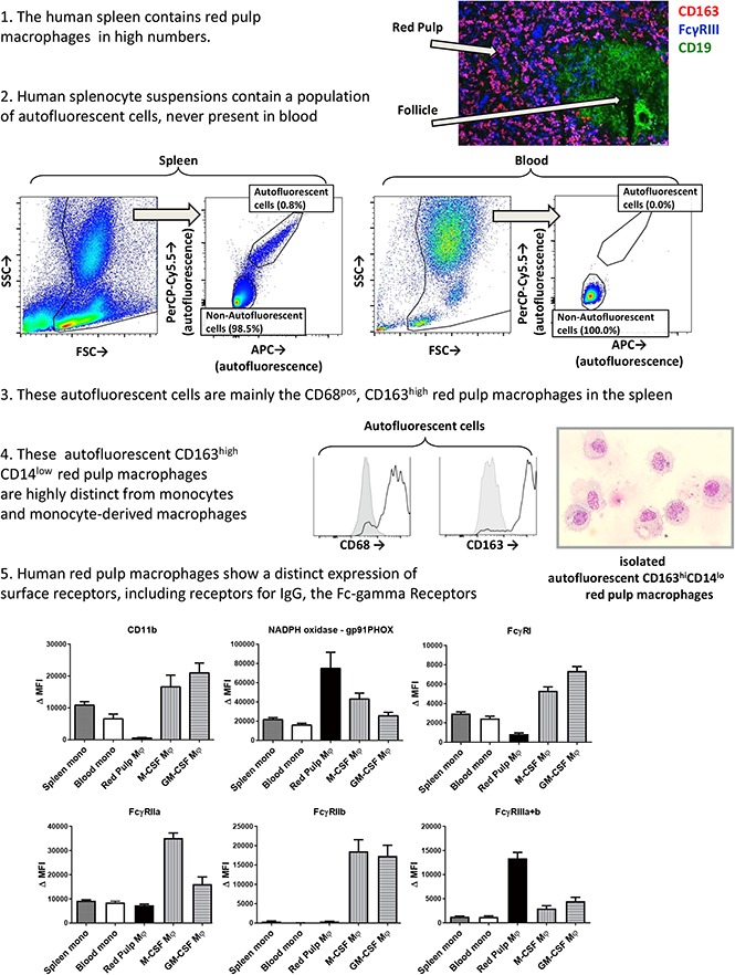
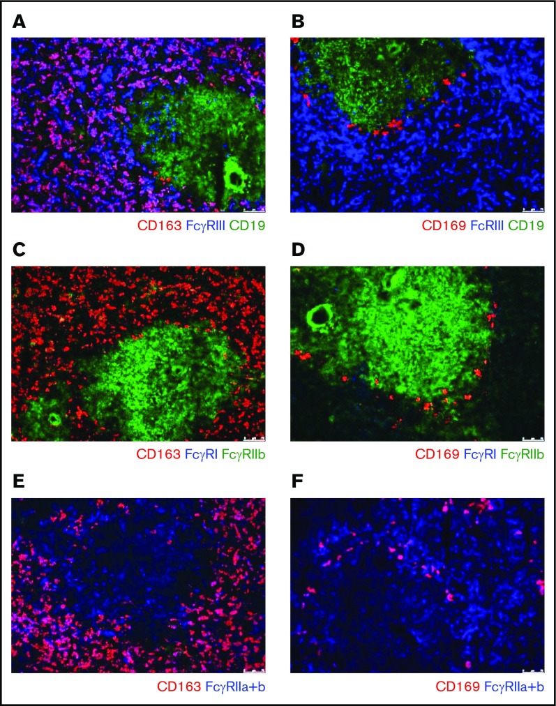
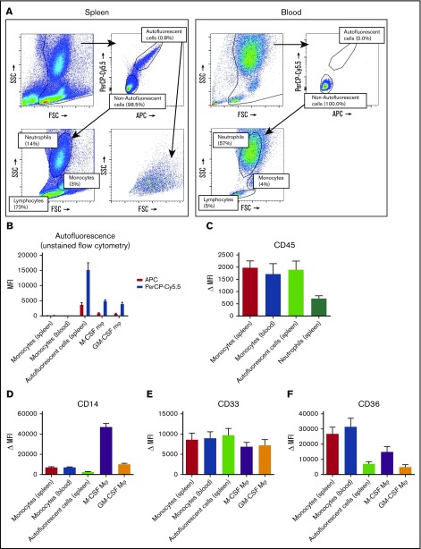
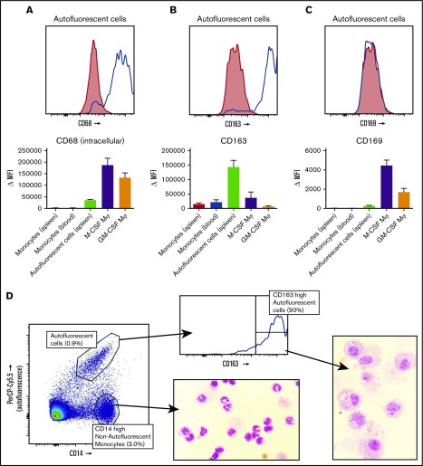
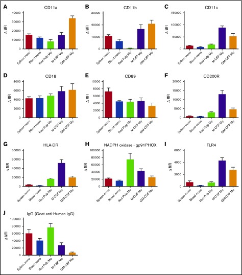
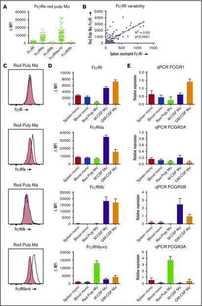
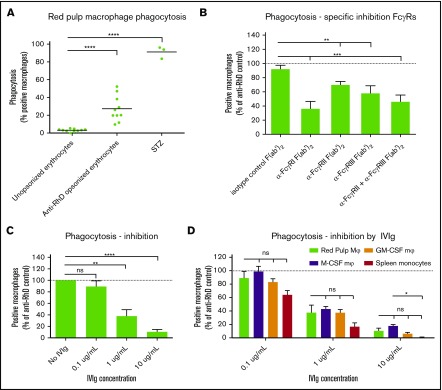
Tissue-resident macrophages in the spleen play a major role in the clearance of immunoglobulin G (IgG)-opsonized blood cells, as occurs in immune thrombocytopenia (ITP) and autoimmune hemolytic anemia (AIHA). Blood cells are phagocytosed via the Fc-γ receptors (FcγRs), but little is known about the FcγR expression on splenic red pulp macrophages in humans, with only a few previous studies that showed conflicting results. We developed a novel method to specifically isolate red pulp macrophages from 82 human spleens. Surface expression of various receptors and phagocytic capacity was analyzed by flow cytometry and immunofluorescence of tissue sections. Red pulp macrophages were distinct from splenic monocytes and blood monocyte-derived macrophages on various surface markers. Human red pulp macrophages predominantly expressed the low-affinity receptors FcγRIIa and FcγRIIIa. In contrast to blood monocyte-derived macrophages, red pulp macrophages did not express the inhibitory FcγRIIb. Red pulp macrophages expressed very low levels of the high-affinity receptor FcγRI. Messenger RNA transcript analysis confirmed this expression pattern. Unexpectedly and despite these differences in FcγR expression, phagocytosis of IgG-opsonized blood cells by red pulp macrophages was dependent on the same FcγRs as phagocytosis by blood monocyte-derived macrophages, especially in regarding the response to IV immunoglobulin. Concluding, we show the distinct nature of splenic red pulp macrophages in human subjects. Knowledge on the FcγR expression and usage of these cells is important for understanding and improving treatment strategies for autoimmune diseases such as ITP and AIHA.


Conflict of interest statement

Conflict-of-interest disclosure: The authors declare no competing financial interest.

Figures

None
Graphical abstract
Figure 1.
Figure 1.
Immunofluorescence of spleen tissue shows FcγR expression pattern of splenic macrophages. Immunofluorescent costainings of (A) CD163, FcγRIII, and CD19; (B) CD169, FcγRIII, and CD19; (C) CD163, FcγRI, and FcγRIIb; (D) CD169, FcγRI, and FcγRIIb; (E) CD163 and FcγRIIa+b; and (F) CD169 and FcγRIIa+b. Original magnification ×10. CD163 marks the red pulp macrophages, CD169 marks the perifollicular zone macrophages, CD19 marks B cells in the follicles. B cells in the follicle are also positive for FcγRIIb. Figures are representative of n = 3 spleens from different donors.
Figure 2.
Figure 2.
Splenocytes contain an autofluorescent cell population of the monocyte/macrophage lineage. (A) Representative flow cytometry plots of unstained single-cell suspensions of splenocytes (left) and blood leukocytes (right) showing an autofluorescent cell population in splenocytes but not in blood. Another consistent feature of splenocytes is the large proportion of lymphocytes, which are in part larger than blood lymphocytes. Percentages are indicated, figure is representative of n = 82 spleens. (B) MFIs for allophycocyanin (APC) and peridinin-chlorophyll protein-Cy5.5 (PerCP-Cy5.5) in unstained samples, comparing the autofluorescent cells in the spleen and spleen monocytes gated as in panel A, with blood monocytes, M-CSF Mφ, and GM-CSF Mφ cultured monocyte-derived macrophages. (C-F) Stainings of CD45, CD14, CD33, and CD36 on various cell types in spleen and blood. Mean ± standard error of the mean (SEM) of n ≥ 8 are shown for each group. FSC, forward scatter; MFI, median fluorescence intensity; Δ MFI, MFI corrected for staining with an isotype control; M-CSF Mφ , monocyte-derived macrophages cultured for 9 days with M-CSF; GM-CSF Mφ, monocyte-derived macrophages cultured for 9 days with GM-CSF; SSC, side scatter.
Figure 3.
Figure 3.
Spleen autofluorescent cells are macrophages and represent the red pulp macrophages of the spleen. Flow cytometry stainings on monocytes in the spleen, blood monocytes, spleen autofluorescent cells, M-CSF, and GM-CSF cultured monocyte-derived macrophages. (A-C, top) Representative histogram on spleen autofluorescent cells (blue line, specific staining; red shading, isotype control). (A-C, bottom) Summary of Δ MFIs of multiple stainings. (A) Intracellular staining for CD68. (B) CD163 staining. (C) CD169 staining. (D) Sorting strategy for splenocytes sorted into autofluorescent CD163highCD14low red pulp macrophages and nonautofluorescent CD163intCD14high spleen monocytes, and May-Grünwald-Giemsa stainings (original magnification ×40) of the sorted cell populations, representative of n = 8 experiments. Mean ± SEM of n ≥ 7 are shown for each group.
Figure 4.
Figure 4.
Red pulp macrophages exhibit an expression pattern distinct from monocytes and monocyte-derived macrophages. Flow cytometry stainings on nonautofluorescent CD163intCD14high spleen monocytes, CD14high blood monocytes gated on FSC/SSC patterns, autofluorescent CD163highCD14low red pulp macrophages from spleen, and M-CSF and GM-CSF cultured monocyte-derived macrophages, (A) CD11a, (B) CD11b, (C) CD11c, (D) CD18, (E) CD89, (F) CD200R, (G) HLA-DR, (H) gp91PHOX (reduced NAD phosphate [NADPH] oxidase complex), (I) TLR4, and (J) IgG (polyclonal goat anti-human IgG). Mean ± SEM of n ≥ 8 are shown for each group.
Figure 5.
Figure 5.
FcγR expression on red pulp macrophages is distinct from the expression on monocytes and monocyte-derived macrophages. (A) Overview of expression of the FcγR isoforms on autofluorescent CD163highCD14low red pulp macrophages from spleen, showing individual values. FcγRI (MoAb 10.1), n = 82; FcγRIIa (MoAb IV.3), n = 57; FcγRIIb (MoAb 2B6), n = 43; FcγRIIIa+b (MoAb 3G8), n = 82; FcγRIIIb (MoAb 5D7), n = 7. (B) Correlation of FcγRI expression on red pulp Mφ with FcγRI levels on neutrophils in the same spleen (n = 82). (C) Representative histograms on autofluorescent CD163highCD14low red pulp macrophages from spleen for FcγRI, FcγRIIa, FcγRIIb, and FcγRIIIa+b (blue line: specific staining, red shading: isotype control). (D) Comparison of flow cytometry stainings on nonautofluorescent CD163intCD14high spleen monocytes, CD14high blood monocytes gated on FSC/SSC pattern, autofluorescent CD163highCD14low red pulp macrophages from spleen, and M-CSF and GM-CSF cultured monocyte-derived macrophages. FcγRI: n ≥ 24 for each group. FcγRIIa: n ≥ 10 for each group. FcγRIIb: n ≥ 15 for each group. To ensure specificity for FcγRIIb, only samples from individuals that cannot express FcγRIIc and with wild-type FCGR2B promoters are presented. FcγRIIIa+b: n ≥ 23 for each group. Mean ± SEM are shown for each group. (E) Quantitative mRNA analysis of various FCGR transcripts encoding FcγRs. The relative expression compared with the expression in pooled whole blood leukocytes, corrected for housekeeping genes, is shown for each transcript. Mean ± SEM of n = 3 are shown for each group.
Figure 6.
Figure 6.
Red pulp macrophages phagocytose IgG-opsonized erythrocytes. (A) Sorted red pulp macrophages phagocytose erythrocytes opsonized with human anti-RhD antibodies, but do not phagocytose unopsonized erythrocytes. Individual values are shown for n = 10 experiments. Serum treated zymosan (STZ) serves as a positive control indicating phagocytic capacity of these cells (n = 3). (B) Phagocytosis of anti-RhD opsonized erythrocytes by red pulp macrophages can be inhibited by F(ab′)2 fragment against specific FcγRs. Isotype control, n = 9; α-FcγRI; n = 3; α-FcγRII, n = 10; α-FcγRIII, n = 10; α-FcγRII+III, n = 9. Data are normalized against phagocytosis by unblocked macrophages. (C) Phagocytosis of anti-RhD opsonized erythrocytes by red pulp macrophages can be inhibited by IVIg in a concentration-dependent manner. n = 4 for each concentration. (D) Comparison of inhibition by IVIg in various cell types. Red pulp macrophages, n = 4; spleen monocytes, n = 3, M-CSF Mφ, n = ≥22; M-CSF, n = 26. Data in panels B-D are normalized against phagocytosis by unblocked macrophages. Mean ± SEM are shown for each group throughout this figure.

References

    1. Crow AR, Lazarus AH. Role of Fcgamma receptors in the pathogenesis and treatment of idiopathic thrombocytopenic purpura. J Pediatr Hematol Oncol. 2003;25(suppl 1):S14-S18. - PubMed
    1. Sandler SG. The spleen and splenectomy in immune (idiopathic) thrombocytopenic purpura. Semin Hematol. 2000;37(1 suppl 1):10-12. - PubMed
    1. Najean Y, Rain JD, Billotey C. The site of destruction of autologous 111In-labelled platelets and the efficiency of splenectomy in children and adults with idiopathic thrombocytopenic purpura: a study of 578 patients with 268 splenectomies. Br J Haematol. 1997;97(3):547-550. - PubMed
    1. Dambuyant C, Thivolet J, Viala JJ, Ville D, Boyer J. Clearance mediated by splenic macrophage membrane receptors for immune complexes in cutaneous vasculitis. J Invest Dermatol. 1982;78(3):194-199. - PubMed
    1. Provan D, Stasi R, Newland AC, et al. International consensus report on the investigation and management of primary immune thrombocytopenia. Blood. 2010;115(2):168-186. - PubMed

Publication types