Review
. 2018 Apr 1;32(7-8):466-478.
doi: 10.1101/gad.309823.117.

Bridging barriers: a comparative look at the blood-brain barrier across organisms

Affiliations
Review

Bridging barriers: a comparative look at the blood-brain barrier across organisms

Natasha M O'Brown et al. Genes Dev. .

Abstract

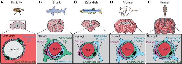
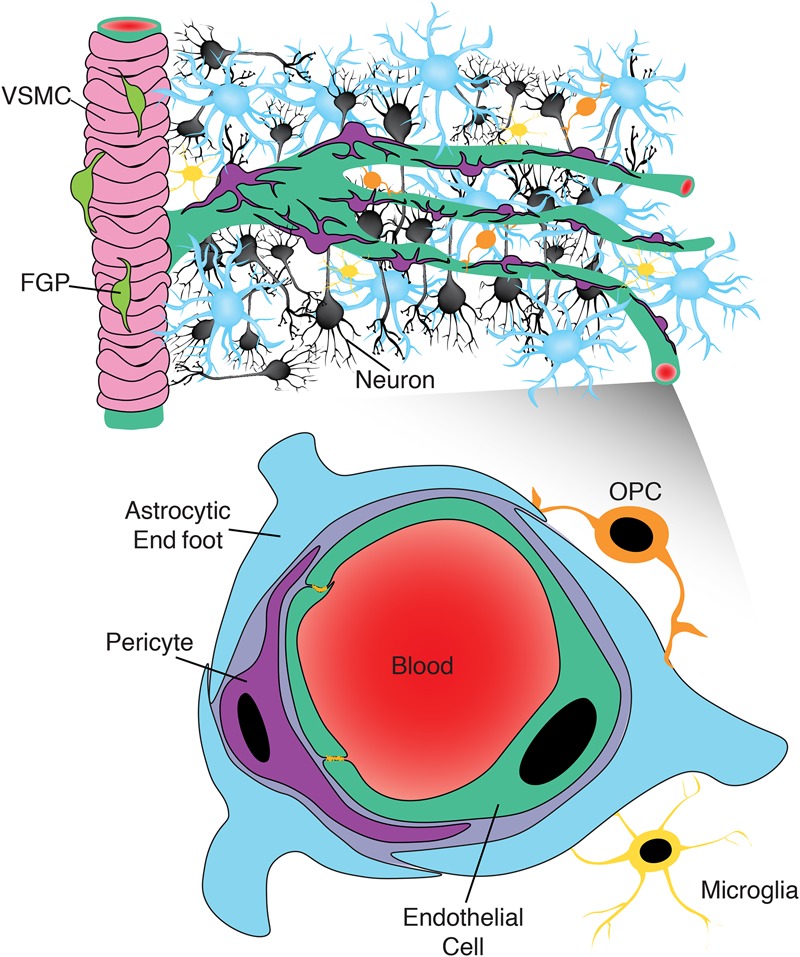
The blood-brain barrier (BBB) restricts free access of molecules between the blood and the brain and is essential for regulating the neural microenvironment. Here, we describe how the BBB was initially characterized and how the current field evaluates barrier properties. We next detail the cellular nature of the BBB and discuss both the conservation and variation of BBB function across taxa. Finally, we examine our current understanding of mouse and zebrafish model systems, as we expect that comparison of the BBB across organisms will provide insight into the human BBB under normal physiological conditions and in neurological diseases.

Keywords: blood–brain barrier; evolution; mouse; zebrafish.


Figures

Figure 1.
Figure 1.
Functional conservation of the barrier across organisms. (A) In fruit flies, the hemolymph–brain barrier partitions the fly neuropil from the hemolymph surrounding the brain, thus performing a function analogous to that of the BBB found in vertebrates. The site of the hemolymph–brain barrier is the SPGs (green), large polyploid cells that enwrap the neural tissue. Subperineurial cells are connected by specialized septate junctions (orange). Perineurial glia (blue) are glial cells that surround the subperineurial layer and also play a role in barrier function. (B) In sharks, the site of the BBB is the perivascular glial cells (green), which are connected by tight junctions (orange) and are found surrounding brain endothelial cells (blue) and pericytes (purple). (C) In zebrafish, the site of the BBB is the capillary endothelial cells (green) that vascularize the brain. These endothelial cells are connected by specialized tight junctions (orange) and are in close contact with brain pericytes (purple). Zebrafish have radial glial cells (blue) that resemble the astrocytes found in mammals; however, their role in the zebrafish BBB has not been well characterized. (D) The site of the mouse BBB is also the capillary endothelial cells (green) that vascularize the brain. As in zebrafish, these cells are connected by specialized tight junctions (orange). BBB endothelial cells together with brain pericytes (purple) and the end feet of astrocytes (blue) comprise the NVU. (E) As in zebrafish and mice, the site of the BBB in humans is also the capillary endothelial cells (green), which are connected by specialized tight junctions (orange). The human NVU also includes pericytes (purple) and astrocytic end feet (blue), which are more prevalent in human brains than in mouse brains.
Figure 2.
Figure 2.
Expanded view of the NVU. While the canonical view of the NVU includes neurons (black), endothelial cells (green), pericytes (purple), and astrocytes (blue), recent evidence suggests that this view may be too limited. Recent work has identified a role for fluorescent granular perithelial cells (FGPs; lime green) in regulating arteriole permeability, marked here by the presence of vascular smooth muscle cells (VSMCs; pink). At the capillary level, there may also be an underappreciated role for cells such as oligodendrocyte precursor cells (OPCs; orange) and microglia (yellow) in regulating the BBB, given their proximity to brain capillaries.

References

    1. Abbott NJ. 2005. Dynamics of CNS barriers: evolution, differentiation, and modulation. Cell Mol Neurobiol 25: 5–23. - PMC - PubMed
    1. Abbott NJ, Rönnbäck L, Hansson E. 2006. Astrocyte–endothelial interactions at the blood–brain barrier. Nat Rev Neurosci 7: 41–53. - PubMed
    1. Adams RA, Bauer J, Flick MJ, Sikorski SL, Nuriel T, Lassmann H, Degen JL, Akassoglou K. 2007. The fibrin-derived γ 377–395 peptide inhibits microglia activation and suppresses relapsing paralysis in central nervous system autoimmune disease. J Exp Med 204: 571–582. - PMC - PubMed
    1. Allsopp G, Gamble HJ. 1979. Light and electron microscopic observations on the development of the blood vascular system of the human brain. J Anat 128: 461–477. - PMC - PubMed
    1. Alvarez JI, Dodelet-Devillers A, Kebir H, Ifergan I, Fabre PJ, Terouz S, Sabbagh M, Wosik K, Bourbonnière L, Bernard M, et al. 2011. The Hedgehog pathway promotes blood–brain barrier integrity and CNS immune quiescence. Science 334: 1727–1731. - PubMed

Publication types