Review
. 2018 Apr 10:12:238.
doi: 10.3389/fnins.2018.00238. eCollection 2018.

The Relationship Between Dopamine Neurotransmitter Dynamics and the Blood-Oxygen-Level-Dependent (BOLD) Signal: A Review of Pharmacological Functional Magnetic Resonance Imaging

Affiliations
Review

The Relationship Between Dopamine Neurotransmitter Dynamics and the Blood-Oxygen-Level-Dependent (BOLD) Signal: A Review of Pharmacological Functional Magnetic Resonance Imaging

Tyler J Bruinsma et al. Front Neurosci. .

Abstract

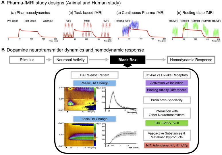
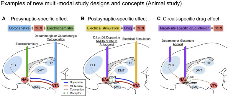
Functional magnetic resonance imaging (fMRI) is widely used in investigations of normal cognition and brain disease and in various clinical applications. Pharmacological fMRI (pharma-fMRI) is a relatively new application, which is being used to elucidate the effects and mechanisms of pharmacological modulation of brain activity. Characterizing the effects of neuropharmacological agents on regional brain activity using fMRI is challenging because drugs modulate neuronal function in a wide variety of ways, including through receptor agonist, antagonist, and neurotransmitter reuptake blocker events. Here we review current knowledge on neurotransmitter-mediated blood-oxygen-level dependent (BOLD) fMRI mechanisms as well as recently updated methodologies aimed at more fully describing the effects of neuropharmacologic agents on the BOLD signal. We limit our discussion to dopaminergic signaling as a useful lens through which to analyze and interpret neurochemical-mediated changes in the hemodynamic BOLD response. We also discuss the need for future studies that use multi-modal approaches to expand the understanding and application of pharma-fMRI.

Keywords: BOLD; dopamine; fMRI; fast-scan cyclic voltammetry; pharma-fMRI.


Figures

Figure 1
Figure 1
(A) Pharma-fMRI study designs. Pharma-fMRI is usually performed under three experimental designs: (a) The red line shows the pharmacodynamics model of a drug (morphine) (Khalili-Mahani et al., 2017). (b) Task-based pharma-fMRI analyzes a behavioral event at baseline and while the drug is under effect (Iannetti and Wise, ; Borsook et al., 2008); (c) continuous collection of fMRI over the course of drug infusion followed by analysis of the change in BOLD signal from pre-drug baseline (Bloom et al., ; De Simoni et al., 2013); and (e) pharmacological resting-state fMRI (pharma-RSfMRI) which examines several short resting-state intervals over the course of drug administration and compares network property changes across different phases of the pharmacokinetic profile (Khalili-Mahani et al., ; Lu and Stein, 2014). Diagram reprinted with permission from Khalili-Mahani et al. (2017). (B) DA neurotransmitter dynamics and hemodynamic response. A schematic of showing the possible factors affecting the link between DA neurotransmitter dynamics and the hemodynamic response (Concept adopted from Jenkins, 2012). Since there are no known voltage-gated vascular receptors, the mechanism by which neurotransmitter release and uptake leads to signaling and release of vasoactive molecules remains unknown, noted here as the “black box” (Jenkins, 2012). Recent updates on DA pathway mechanisms demonstrate a complex release pattern having both phasic and tonic states, dynamically modifying the basal tone of neuronal activity, as seen in disease or drug-induced states (Owesson-White et al., ; Grace, 2016). The phasic DA response in the upper left color map is from electrically stimulating (2 s) the nigrostriatal pathway and conducting neurochemical analysis in the caudate, which was confirmed by fMRI in a within subject study of a non-human primate (Min et al., 2016). Each electrochemical signal was converted to molar (M) concentration showing the difference from baseline to an electrically evoked response. The tonic DA response in the lower left color map is from systematic administration of nomifensine, a DA transporter reuptake blocker, and recording the electrochemical response in striatum over a time period of 2 h in rodents (Oh et al., 2016). Other factors are discussed in the current review, including DA receptor family type specific effects, brain area specificity, and DA integration with other neurotransmitters. Finally, neuronal activity would influence vasoactive substances (NO, K+) and metabolic by-products (Adenosine, H+, CO2) affecting the local hemodynamic response.
Figure 2
Figure 2
Examples of new multi-modal study designs and concepts in large animal (swine) brain diagram: (A) Presynaptic-specific effect. Optogenetics enables cell-type specific stimulation, and the fMRI signal induced by stimulation would provide insight into presynaptic-specific effects on BOLD (Albaugh et al., ; Helbing et al., ; Lohani et al., 2017) along with electrochemical information; (B) Postsynaptic-specific effect. Electrical stimulation of a specific pathway forces non-specific activity to the neuronal circuit involving all cell-types near the electrode (Kringelbach et al., 2007), combined with electrode-metal induced susceptibility artifact corrected fMRI (In et al., 2017). Administering receptor antagonists along the stimulated pathway enables the evaluation of receptor-specific effects on BOLD (Ross et al., 2016); (C) Circuit-specific drug effect. Popular in animal studies, brain circuit-specific intracranial drug injection (Wise and Hoffman, ; Kim et al., 2014) combined with fMRI, could open new possibilities for studying circuit-specific neurotransmitter (agonist) effects on BOLD by limiting the involvement of presynaptic dynamics.

References

    1. Adamantidis A. R., Tsai H. C., Boutrel B., Zhang F., Stuber G. D., Budygin E. A., et al. (2011). Optogenetic interrogation of dopaminergic modulation of the multiple phases of reward-seeking behavior. J. Neurosci. 31, 10829–10835. 10.1523/JNEUROSCI.2246-11.2011 - DOI - PMC - PubMed
    1. Albaugh D. L., Salzwedel A., Van Den Berge N., Gao W., Stuber G. D., Shih Y. Y. (2016). Functional magnetic resonance imaging of electrical and optogenetic deep brain stimulation at the rat nucleus accumbens. Sci. Rep. 6:31613. 10.1038/srep31613 - DOI - PMC - PubMed
    1. Angelovski G., Tóth É. (2017). Strategies for sensing neurotransmitters with responsive MRI contrast agents. Chem. Soc. Rev. 46, 324–336. 10.1039/C6CS00154H - DOI - PubMed
    1. Atcherley C. W., Laude N. D., Parent K. L., Heien M. L. (2013). Fast-scan controlled-adsorption voltammetry for the quantification of absolute concentrations and adsorption dynamics. Langmuir 29, 14885–14892. 10.1021/la402686s - DOI - PubMed
    1. Atcherley C. W., Wood K. M., Parent K. L., Hashemi P., Heien M. L. (2015). The coaction of tonic and phasic dopamine dynamics. Chem. Commun. 51, 2235–2238. 10.1039/C4CC06165A - DOI - PMC - PubMed

LinkOut - more resources