. 2018 Apr 10:9:658.
doi: 10.3389/fimmu.2018.00658. eCollection 2018.

Interleukin-15-Cultured Dendritic Cells Enhance Anti-Tumor Gamma Delta T Cell Functions through IL-15 Secretion

Affiliations

Interleukin-15-Cultured Dendritic Cells Enhance Anti-Tumor Gamma Delta T Cell Functions through IL-15 Secretion

Heleen H Van Acker et al. Front Immunol. .

Abstract

Dendritic cell (DC) vaccination can be an effective post-remission therapy for acute myeloid leukemia (AML). Yet, current DC vaccines do not encompass the ideal stimulatory triggers for innate gamma delta (γδ) T cell anti-tumor activity. Promoting type 1 cytotoxic γδ T cells in patients with AML is, however, most interesting, considering these unconventional T cells are primed for rapid function and exert meaningful control over AML. In this work, we demonstrate that interleukin (IL)-15 DCs have the capacity to enhance the anti-tumoral functions of γδ T cells. IL-15 DCs of healthy donors and of AML patients in remission induce the upregulation of cytotoxicity-associated and co-stimulatory molecules on the γδ T cell surface, but not of co-inhibitory molecules, incite γδ T cell proliferation and stimulate their interferon-γ production in the presence of blood cancer cells and phosphoantigens. Moreover, the innate cytotoxic capacity of γδ T cells is significantly enhanced upon interaction with IL-15 DCs, both towards leukemic cell lines and allogeneic primary AML blasts. Finally, we address soluble IL-15 secreted by IL-15 DCs as the main mechanism behind the IL-15 DC-mediated γδ T cell activation. These results indicate that the application of IL-15-secreting DC subsets could render DC-based anti-cancer vaccines more effective through, among others, the involvement of γδ T cells in the anti-leukemic immune response.

Keywords: acute myeloid leukemia; dendritic cell vaccination; immunotherapy; interleukin-15; γδ T cells.


Figures

Figure 1
Figure 1
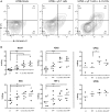
IL-15 dendritic cells (DCs) induce γδ T cell proliferation in a contact-independent manner. (A) γδ T cells in full peripheral blood mononuclear cell (PBMC) fraction were stimulated with isopentenyl pyrophosphate (IPP), IL-15 DCs (E:T ratio = 1:10) or IL-15 DCs + IPP for 5 days. Unstimulated PBMCs (−) were used as negative control. γδ T cell proliferation, as assessed by reduction in CFSE staining, was determined by flow cytometry. The percentage of cells to have undergone at least one cell division is shown (n = 12). One-way ANOVA with Bonferroni’s Multiple Comparison Test. (B) The histogram overlay shows CFSE dilution of viable γδ T cells within unstimulated PBMCs (filled gray), PBMCs stimulated with IPP (dotted line), IL-15 DCs (dashed line), and IL–15 DCs + IPP (full line) for one representative donor. (C) Dot plots of one representative donor (n = 3) showing the CFSE-dilution of proliferated γδ T cells in PBMCs stimulated with IL-15 DCs + IPP with and without IL-15 DCs in transwell (TW). (D) γδ T cell proliferation over time (experimental setup in line with panel A; n = 3). *** p < 0.001.
Figure 2
Figure 2
IL-15 dendritic cells (DCs) stimulate γδ T cell IFN-γ production in the presence of a leukemic environment through a contact-independent mechanism (A) γδ T cells in full peripheral blood mononuclear cell fraction were examined for IFN-γ production by intracellular staining after overnight stimulation with isopentenyl pyrophosphate (IPP), IL-15 DCs (E:T ratio = 1:10), tumor cells (E:T ratio = 1:10), and all possible combinations. Resting γδ T cells (−) were used as negative point of reference. Data are depicted as the mean of ten independent donors. One-way ANOVA with Bonferroni’s Multiple Comparison Test. (B) Representative FACS dot plots of one healthy donor out of 10 showing IFN-γ production by γδ T cells. (C) In parallel, the same experiment was carried out except that IL-15 DCs were separated from the cultures by 0.4-µm transwell (TW) inserts (n = 3). *** p < 0.001, ** p < 0.01, and * p < 0.5.
Figure 3
Figure 3
IL-15, secreted by IL-15 dendritic cells (DCs), provides an important signal for DC-mediated γδ T cell proliferation and IFN-γ production. (A) Representation of the IL-15 secretion level (pg/mL), as determined by Meso Scale Discovery immunoassay, in 48-hour wash-out supernatant of IL-15 DC cultures (1 × 106 cells/mL; n = 3). (B) CFSE-labeled peripheral blood mononuclear cells (PBMCs) were co-cultured with 1 ng/mL recombinant IL-15 or IL-15 DCs (E:T ratio = 1:10). In certain conditions, neutralizing IL-15 monoclonal antibodies (mAbs) (aIL-15) or isotype control mAbs (IgG1) were added to the IL-15 DC cultures 1 hour prior to the addition of PBMCs. Percentages refer to the proportion of γδ T cells that proliferated within 5 days (n = 6). Friedman test with Dunn’s Multiple Comparison Test. (C) After overnight culture of γδ T cells in full PBMC fraction with IL-15 DCs + IPP (= cond) and K562 or Daudi tumor cells (E:T ratio = 1:10), with anti-IL-15 mAbs (aIL-15) or with isotype control mAbs (IgG1), intracellular IFN-γ production was measured with flow cytometry in γδ T cells (n = 6). Friedman test with Dunn’s Multiple Comparison Test. ** p < 0.01 and * p < 0.5; ns, p > 0.5.
Figure 4
Figure 4
The cytotoxic capacity of γδ T cells is radically increased by IL-15 dendritic cells (DCs). (A) Representative example of primary acute myeloid leukemia blast killing (UPNb) from one out of three donors. (B) Cytotoxicity was determined of γδ cell ± IL-15 DC (co-)cultures with or without isopentenyl pyrophosphate after 24–36 hours. Unstimulated γδ T cells (−) were used to define basal killing capacity. Target cells were added at an E:T ratio of 2:1. Percentage tumor cell killing was ascertained by Annexin-V/PI staining after 4 hours and calculated using the formula specified in Section “Materials and Methods.” Donors are represented by unique symbols. Friedman test with Dunn’s Multiple Comparison Test. *** p < 0.001, ** p < 0.01, and * p < 0.5.
Figure 5
Figure 5
Functional activation of γδ T cells of acute myeloid leukemia (AML) patients by IL-15 dendritic cells (DCs). (A) Bar graphs depict γδ T cell proliferation upon 5-day culture (−) with isopentenyl pyrophosphate (IPP), IL-15 DCs (E:T ratio = 1:10) or IL-15 DCs + IPP. (B) Percentage IFN-γ-positive γδ T cells after overnight culture (−) with IPP, IL-15 DCs (E:T ratio = 1:10), and/or tumor cells (E:T ratio = 1:10) measured by flow cytometry for four AML patients in first complete remission. The IFN-γ production by γδ T cells of unique patient number (UPN)1&2 and UPN3&4 is shown separately, as they differ from each other in response to stimuli. Blood from UPN1&2 and UPN3&4 was drawn after termination of induction and consolidation chemotherapy (I + C) and induction chemotherapy (I), respectively. No statistics were performed due to limited numbers.

Similar articles

Cited by

References

    1. Dohner H, Weisdorf DJ, Bloomfield CD. Acute myeloid leukemia. N Engl J Med (2015) 373(12):1136–52. 10.1056/NEJMra1406184 - DOI - PubMed
    1. De Kouchkovsky I, Abdul-Hay M. Acute myeloid leukemia: a comprehensive review and 2016 update. Blood Cancer J (2016) 6(7):e441. 10.1038/bcj.2016.50 - DOI - PMC - PubMed
    1. Martner A, Thoren FB, Aurelius J, Hellstrand K. Immunotherapeutic strategies for relapse control in acute myeloid leukemia. Blood Rev (2013) 27(5):209–16. 10.1016/j.blre.2013.06.006 - DOI - PubMed
    1. Hirsch P, Tang R, Abermil N, Flandrin P, Moatti H, Favale F, et al. Precision and prognostic value of clone-specific minimal residual disease in acute myeloid leukemia. Haematologica (2017) 102(7):1227–37. 10.3324/haematol.2016.159681 - DOI - PMC - PubMed
    1. Schurch CM, Riether C, Ochsenbein AF. Dendritic cell-based immunotherapy for myeloid leukemias. Front Immunol (2013) 4:496. 10.3389/fimmu.2013.00496 - DOI - PMC - PubMed

Publication types