Review
. 2018 Mar 5:2018:6436453.
doi: 10.1155/2018/6436453. eCollection 2018.

Regulation of Central Nervous System Myelination in Higher Brain Functions

Affiliations
Review

Regulation of Central Nervous System Myelination in Higher Brain Functions

Mara Nickel et al. Neural Plast. .

Abstract

The hippocampus and the prefrontal cortex are interconnected brain regions, playing central roles in higher brain functions, including learning and memory, planning complex cognitive behavior, and moderating social behavior. The axons in these regions continue to be myelinated into adulthood in humans, which coincides with maturation of personality and decision-making. Myelin consists of dense layers of lipid membranes wrapping around the axons to provide electrical insulation and trophic support and can profoundly affect neural circuit computation. Recent studies have revealed that long-lasting changes of myelination can be induced in these brain regions by experience, such as social isolation, stress, and alcohol abuse, as well as by neurological and psychiatric abnormalities. However, the mechanism and function of these changes remain poorly understood. Myelin regulation represents a new form of neural plasticity. Some progress has been made to provide new mechanistic insights into activity-independent and activity-dependent regulations of myelination in different experimental systems. More extensive investigations are needed in this important but underexplored research field, in order to shed light on how higher brain functions and myelination interplay in the hippocampus and prefrontal cortex.


Figures
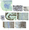
Figure 1
Figure 1
Myelin formation in the hippocampus of humans at different developmental stages. (a) At the 37th gestational week (GW), MBP-positive myelin segments (brown) are present in the fimbria (F), in the entire alveus (A), and in the stratum lacunosum-moleculare (L-M) of the CA1 region. MBP-positive OL cell bodies (arrowheads) and myelin segments (arrows) are present in the fimbria fornicis (b), in the alveus (c), and in the stratum lacunosum-moleculare (L-M) (d) in an infant born at the 37th GW. (e) At 2 years of age, strong MBP staining was observed in the alveus, in the stratum lacunosum-moleculare (L-M) of Ammon's horn, and in the outer half of the molecular layer (M) of the dentate gyrus. MBP-positive myelin segments are also present in the strata oriens (O), pyramidale (P), and radiatum (R) of Ammon's horn. (f) MBP-positive myelin segments in the hilus (H) of dentate gyrus at 2 years of age. (g) MBP-positive myelin segments start to form a dense network in the hilus of the dentate gyrus at the age of 11 years. (h) Much denser network of myelin segments in the hilus of a 53-year-old adult. Scale bars: 1000 μm in (e), 500 μm in (a), 250 μm in (g) and (h), and 100 μm in (b)–(d) and (f). This figure is modified from Abrahám et al. [31], with copyright permission for reusing the figure panels.
Figure 2
Figure 2
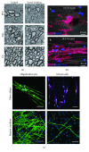
Axonal activity-dependent and activity-independent regulation of myelination. (a) Prolonged social isolation of adult mice induces hypomyelination in the PFC. Electron micrographs of axons in the PFC, nucleus accumbens (NAc), and corpus callosum (CC) from control and isolated mice (modified from Liu et al. [47], with copyright permission for reusing the figure panels). (b) The fiber diameter is sufficient to initiate myelination in a neuron-free culture. To determine the minimum fiber diameter at which oligodendrocytes commence wrapping in our system, Lee et al. analyzed oligodendrocytes cultured on nanofibers ranging from 0.2 to 4.0 μm in diameter and quantified the total number of MBP+ segments normalized to the fiber distribution on each coverslip. The minimum fiber diameter threshold for oligodendrocyte myelination is approximately 0.4 μm, which is supported by immunostaining of cultures for MBP and DAPI in the presence of small-diameter (A, 0.2–0.4 μm) and large-diameter (B, 2.0–4.0 μm) fibers. Electron-spun polystyrene or poly-L-lactic acid (PLLA) nanofibers with diameters ranging from 0.2 μm to 4.0 μm were engineered (modified from [62] with copyright permission for reusing the figure panels). (c) OLs have the unique, intrinsic capability to generate compact membrane sheaths and physiological internode lengths on microfibers. Confocal stacks of rat primary cortical OLs or Schwann cells cultured 14 or 21 days, respectively, on 1-2 μm microfibers or neurons (green: MBP, blue: Hoechst, and purple: S-100) (modified from Bechler et al. [64], with copyright permission for reusing the figure panels). Scale bars: 0.5 μm in (a) and 40 μm in (b) and (c).
Figure 3
Figure 3
Mechanisms underlying activity-dependent regulation of myelination. (a) Diagram of the multiple steps involved in the development of OLs. OPCs differentiate into multipolar premyelinating OLs, which mature into myelinating OLs. Mature OLs form myelin segments on multiple axons simultaneously. Regulation can occur at different steps during development, eventually leading to altered myelin formation. (b) Nascent myelin sheaths are stabilized by activity-dependent secretion. Representative confocal images show the retraction of existing sheaths during 15 h time-lapse imaging in sibling control (left) and Tg(neurod1:TeNT-EGFP) larvae (right). In the right panel, expression of TeNT-EGFP disrupted axonal activity-dependent secretion. Images are lateral views of the dorsal spinal cord, and the time relative to the start of image acquisition is indicated for each image. For demonstrative purposes, sheaths stable for the entire time-lapse are shaded in blue. Retracting sheaths are shaded in red and are also indicated by red arrowheads (modified from Hines et al. [71], with copyright permission for reusing the figure panels). Scale bar: 5 μm.

Similar articles

Cited by

References

    1. Preston A. R., Eichenbaum H. Interplay of hippocampus and prefrontal cortex in memory. Current Biology. 2013;23(17):R764–R773. doi: 10.1016/j.cub.2013.05.041. - DOI - PMC - PubMed
    1. Frankland P. W., Bontempi B. The organization of recent and remote memories. Nature Reviews Neuroscience. 2005;6(2):119–130. doi: 10.1038/nrn1607. - DOI - PubMed
    1. Kitamura T., Ogawa S. K., Roy D. S., et al. Engrams and circuits crucial for systems consolidation of a memory. Science. 2017;356(6333):73–78. doi: 10.1126/science.aam6808. - DOI - PMC - PubMed
    1. Siapas A. G., Lubenov E. V., Wilson M. A. Prefrontal phase locking to hippocampal theta oscillations. Neuron. 2005;46(1):141–151. doi: 10.1016/j.neuron.2005.02.028. - DOI - PubMed
    1. Benchenane K., Peyrache A., Khamassi M., et al. Coherent theta oscillations and reorganization of spike timing in the hippocampal- prefrontal network upon learning. Neuron. 2010;66(6):921–936. doi: 10.1016/j.neuron.2010.05.013. - DOI - PubMed

Publication types

LinkOut - more resources