. 2018 Mar 6:2018:8478943.
doi: 10.1155/2018/8478943. eCollection 2018.

Emblic Leafflower (Phyllanthus emblica L.) Fruits Ameliorate Vascular Smooth Muscle Cell Dysfunction in Hyperglycemia: An Underlying Mechanism Involved in Ellagitannin Metabolite Urolithin A

Affiliations

Emblic Leafflower (Phyllanthus emblica L.) Fruits Ameliorate Vascular Smooth Muscle Cell Dysfunction in Hyperglycemia: An Underlying Mechanism Involved in Ellagitannin Metabolite Urolithin A

Junxuan Zhou et al. Evid Based Complement Alternat Med. .

Abstract

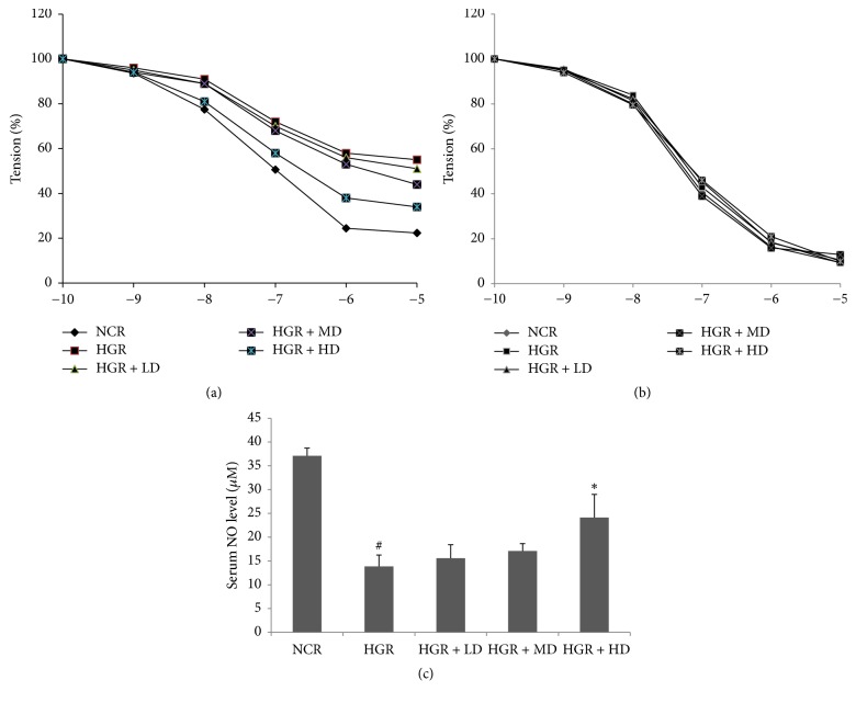
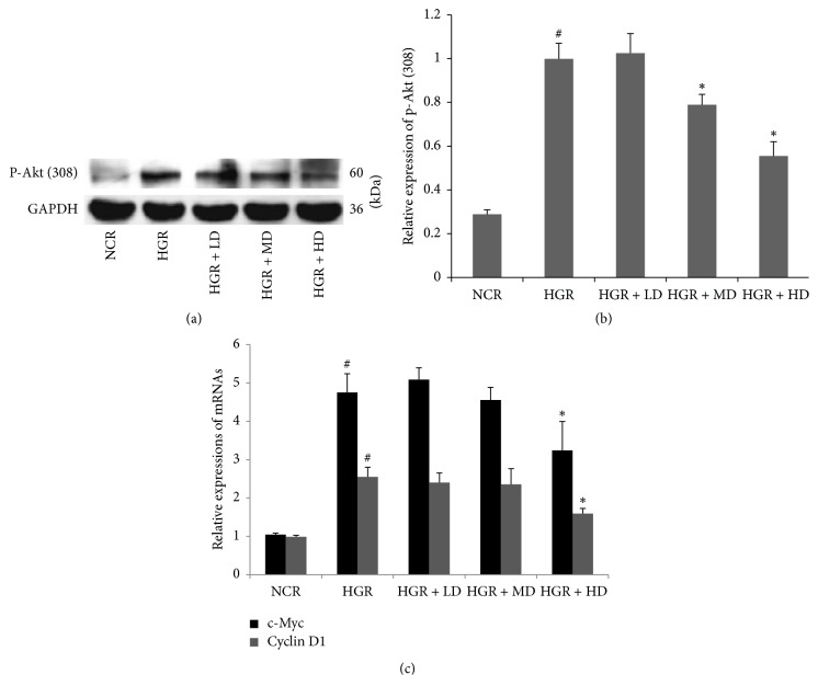
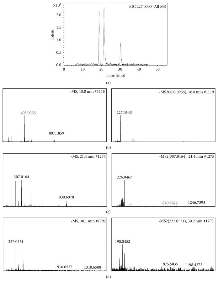
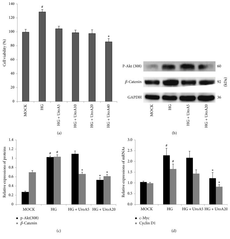
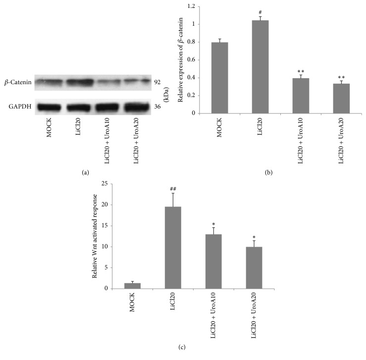
Ellagitannins in Phyllanthus emblica L. (emblic leafflower fruits) have been thought of as the beneficial constituents for ameliorating endocrinal and metabolic diseases including diabetes. However, the effect of emblic leafflower fruits on diabetic vascular complications involved in ellagitannin-derived urolithin metabolites is still rare. In this study, acetylcholine-induced endothelium-independent relaxation in aortas was facilitated upon emblic leafflower fruit consumption in the single dose streptozotocin-induced hyperglycemic rats. Emblic leafflower fruit consumption also suppressed the phosphorylation of Akt (Thr308) in the hyperglycemic aortas. More importantly, urolithin A (UroA) and its derived phase II metabolites were identified as the metabolites upon emblic leafflower fruit consumption by HPLC-ESI-Q-TOF-MS. Moreover, UroA reduced the protein expressions of phosphor-Akt (Thr308) and β-catenin in a high glucose-induced A7r5 vascular smooth muscle cell proliferation model. Furthermore, accumulation of β-catenin protein and activation of Wnt signaling in LiCl-triggered A7r5 cells were also ameliorated by UroA treatment. In conclusion, our data demonstrate that emblic leafflower fruit consumption facilitates the vascular function in hyperglycemic rats by regulating Akt/β-catenin signaling, and the effects are potentially mediated by the ellagitannin metabolite urolithin A.


Figures

Figure 1
Figure 1
Emblic leafflower fruit (ELF) consumption ameliorates endothelium-dependent vasodilatation in hyperglycemic rats. (a) Acetylcholine-induced endothelium-dependent relaxation (EDR), (b) SNP-induced endothelium-independent relaxation (EIR) in rat aortic rings, and (c) serum nitric oxide (NO) levels in (1) normal control rats (NCR); (2) hyperglycemic rats (HGR); (3) hyperglycemic rats treated with low dose of ELF (LD, ELF 25 g/kg); (4) hyperglycemic rats treated with median dose (MD, ELF 50 g/kg); (5) hyperglycemic rats treated with high dose (HD, ELF 75 g/kg). Mean ± SD, n = 6. #P < 0.05 versus NCR; P < 0.05 versus HGR.
Figure 2
Figure 2
Emblic leafflower fruit (ELF) administration suppresses the protein expression of phosphorylated-Akt (Thr308) and the mRNA transcriptions of c-Myc and cyclin D1. The protein expression of phosphorylated-Akt (Thr308) (a, b) and the mRNA transcriptions of c-Myc and cyclin D1 (c) in rat aortic rings in (1) normal control rats (NCR); (2) hyperglycemic rats (HGR); (3) hyperglycemic rats treated with low dose of ELF (LD, ELF 25 g/kg); (4) hyperglycemic rats treated with median dose (MD, ELF 50 g/kg); (5) hyperglycemic rats treated with high dose (HD, ELF 75 g/kg). n = 3. #P < 0.05 versus NCR; P < 0.05 versus HGR.
Figure 3
Figure 3
Urolithin A (UroA) related metabolites were converted from ellagitannins in emblic leafflower fruits in vivo. The urine samples in hyperglycemic rats treated with high dose of ELF were collected before sacrifice and further analyzed by HPLC-ESI-Q-TOF-MS. The peaks involved in UroA (m/z 227.0539[M-H]−) in extracted ion chromatogram (EIC) (a) were identified as UroA glucuronic acid (b), UroA-sulfate (b), and UroA (d), respectively.
Figure 4
Figure 4
UroA attenuates high glucose- (HG-) induced A7r5 cell proliferation by suppressing Akt/β-catenin signaling. (a) The effect of UroA on high glucose- (HG-) induced A7r5 cell proliferation. Cells were seeded in 96 well and preincubated with HG (30 mM) for 12 h. The cells were treated by UroA (5–40 μM). CCK-8 assay was performed to determine cell viability as described in Materials and Methods. (b) Western blotting analysis for the protein expressions of phosphorylated-Akt (Thr308) (60 kD) and β-catenin (92 kD) in HG-incubated A7r5 cells. (C) Histograms show phosphorylated-Akt (Thr308) and β-catenin protein levels quantified by western blot optical analysis. (c) The effect of UroA on the mRNA expressions of c-Myc and cyclin D1 was quantified by qPCR in HG-induced VSMC proliferating model. Quantitative data are mean ± SD, n = 3. #P < 0.05 versus MOCK; P < 0.05 versus HG group.
Figure 5
Figure 5
UroA suppresses LiCl-induced activation of Wnt/β-catenin signaling in A7r5 cells. (a, b) Cells were preincubated with LiCl (20 mM) for 3 h. Then the cells were further treated with UroA for 48 h. Western blotting analysis was applied for quantifying the protein expression of β-catenin (92 kD) in LiCl-pretreated A7r5 cells. (c) UroA suppressed Tcf/Lef-mediated transcriptional activity in A7r5 cells. Cells were transiently cotransfected with TopFlash or FopFlash and Renilla reporter plasmid with Lipofectamine 3000. After posttransfection, cells were treated as described in Materials and Methods. Luciferase values were measured using a dual-luciferase reporter assay system. Quantitative data are mean ± SD, n = 3. #P < 0.05, ##P < 0.01 versus MOCK; P < 0.05 and ∗∗P < 0.01 versus LiCl20 group.

References

    1. Yechoor V., Chan L. Gene therapy progress and prospects: Gene therapy for diabetes mellitus. Gene Therapy. 2005;12(2):101–107. doi: 10.1038/sj.gt.3302412. - DOI - PubMed
    1. Grundy S. M., Benjamin I. J., Burke G. L., et al. Diabetes and cardiovascular disease: a statement for healthcare professionals from the american heart association. Circulation. 1999;100(10):1134–1146. doi: 10.1161/01.CIR.100.10.1134. - DOI - PubMed
    1. Lacigova S., Brozova J., Cechurova D., Tomesova J., Krcma M., Rusavy Z. The influence of cardiovascular autonomic neuropathy on mortality in type 1 diabetic patients; 10-year follow-up. Biomedical Papers. 2016;160(1):111–117. doi: 10.5507/bp.2015.063. - DOI - PubMed
    1. Schalkwijk C. G., Stehouwer C. D. A. Vascular complications in diabetes mellitus: the role of endothelial dysfunction. Clinical Science. 2005;109(2):143–159. doi: 10.1042/CS20050025. - DOI - PubMed
    1. Chen S., Evans T., Mukherjee K., Karmazyn M., Chakrabarti S. Diabetes-induced myocardial structural changes: Role of endothelin-1 and its receptors. Journal of Molecular and Cellular Cardiology. 2000;32(9):1621–1629. doi: 10.1006/jmcc.2000.1197. - DOI - PubMed