. 2018 Mar 6:2018:6075705.
doi: 10.1155/2018/6075705. eCollection 2018.

CT and MRI Findings of Soft Tissue Adult Fibrosarcoma in Extremities

Affiliations

CT and MRI Findings of Soft Tissue Adult Fibrosarcoma in Extremities

Hexiang Wang et al. Biomed Res Int. .

Abstract

Objective: To characterize and evaluate CT and MRI features of extremity soft tissue adult fibrosarcoma.

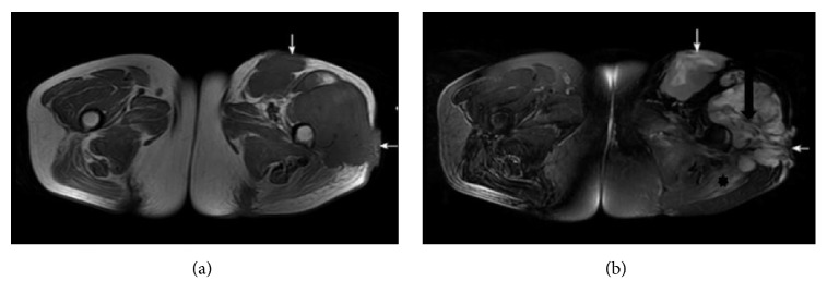
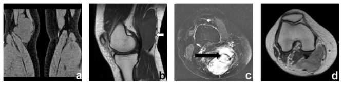
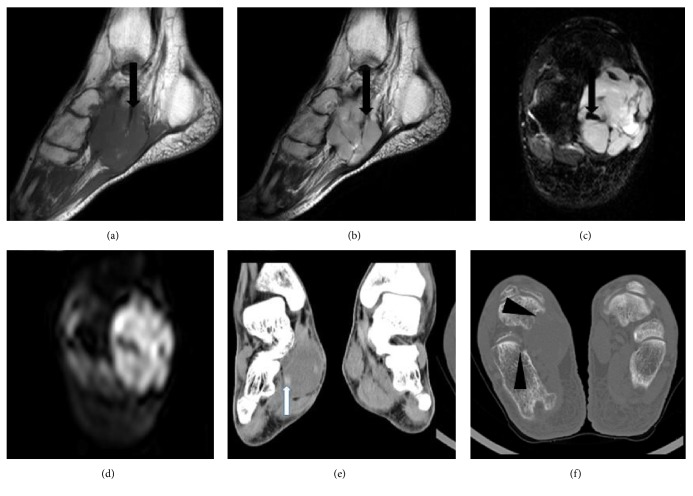
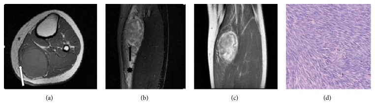
Methods: CT and MRI images from 10 adult patients with pathologically proven fibrosarcomas were retrospectively analyzed with regard to tumor location, size, number, shape, margins, attenuation, signal intensity, and enhancement patterns on MR images. Additionally, the relationships between lesions, deep fascia, and change in adjacent bones were also assessed.

Results: Nineteen tumor lesions in 10 patients were selected for this study. Eighteen lesions were lobulated and one was oval in shape. Most cases were located under the deep fascia, including seven cases that had a nodular lump adjacent to the deep fascia and one case that had broken lesion through the deep fascia. On CT, the adult fibrosarcomas mostly showed iso-attenuated soft tissue masses (n = 6). On MRI, all the cases (n = 9) displayed low signal on T1-weighted imaging (T1WI) and heterogeneous low and high intensity signals on T2-weighted imaging (T2WI), with band-like areas of low signal on both T1WI and T2WI. On contrast-enhanced MRI images, three cases showed heterogeneous peripheral enhancement and one case demonstrated a spoke-wheel-like enhancement. Eight cases showed muscle edema signals in the peritumoral muscle and one case involved adjacent bone.

Conclusion: CT and MR imaging have respective advantages in diagnosing adult fibrosarcoma. Combined application of CT and MR is recommended for patients with suspected adult fibrosarcoma.


Figures

Figure 1
Figure 1
A 43-year-old female with a moderately differentiated adult fibrosarcoma. (a, b) Axial SE T1WI and fat suppressed FSE T2WI, exhibiting two lobulated long T1 mixed T2 signal masses in the left hip deep fascia. The band-like areas of low signal suggest tumor fibrous septa (black arrow in (b)), and the patch-shaped regions of long T1 and T2 signal in the left gluteus maximus muscle represent muscle edema (asterisk in (b)). The lesion broke through the deep fascia (white arrows in (a), (b)).
Figure 2
Figure 2
A 47-year-old female with well-differentiated adult fibrosarcoma. (a) A coronal CT scan exhibited a lobulated iso-attenuating soft tissue mass in the right popliteal fossa. (b) Sagittal T1WI showed the lesion with a nodular lump close to the deep fascia (white arrow). (c) Axial fat suppressed FSE T2WI showed band-like areas of low signal suggesting tumor fibrous septa (black arrow). (d) An axial SE T1W contrast-enhanced scan exhibited heterogeneous peripheral enhancement.
Figure 3
Figure 3
A 46-year-old female with a poorly differentiated adult fibrosarcoma. (a, b) Sagittal T1WI and T2WI exhibited a lobulated long T1 and mixed T2 signal mass in the right plantar deep fascia, with band-like areas of long T1 and short T2 signal (black arrows in (a) and (b)) indicating tumor fibrous septa. (c, d) Axial fat suppressed FSE T2WI and DWI show mixed signals, with more nodular short T2 signals (black arrow in (c)). The high lesion signal on DWI indicated dense cellularity. (e, f) Reformatted coronal images of soft tissue and axial bone images displaying an iso-attenuating soft tissue mass, with a slightly higher density small nodule (white arrow in (e)) and partial bone destruction (arrow heads in (f)).
Figure 4
Figure 4
An 18-year-old female with a moderately differentiated adult fibrosarcoma. (a, b) Axial SE T1WI and coronal fat suppressed FSE T2WI exhibiting lobulated long T1 and mixed T2 signal masses in the left calf deep fascia, with a nodular lump close to the deep fascia (white arrow in (a)). T2WI exhibited band-like areas of low signal (black arrow in (b)) with patch-shaped long T2 signal regions in surrounding muscle, representing muscle edema (asterisk in (b)). (c) Coronal SE T1W enhanced scan showing a spoke-wheel-like enhancement pattern. (d) Pathological image (He ×100), with tumor cells and collagen fibers arranged in crisscrossing bundles, demonstrating a characteristic “herringbone” arrangement.

References

    1. Meyerding H., Broders A., Hargrave R. Clinical aspects of fibrosarcoma of the soft tissues of the extremities. Surgery, Gynecology & Obstetrics. article 62, 1936
    1. Toro J. R., Travis L. B., Hongyu J. W., Zhu K., Fletcher C. D. M., Devesa S. S. Incidence patterns of soft tissue sarcomas, regardless of primary site, in the surveillance, epidemiology and end results program, 1978–2001: an analysis of 26,758 cases. International Journal of Cancer. 2006;119(12):2922–2930. doi: 10.1002/ijc.22239. - DOI - PubMed
    1. Fisher C. The value of electronmicroscopy and immunohistochemistry in the diagnosis of soft tissue sarcomas: a study of 200 cases. Histopathology. 1990;16(5):441–454. doi: 10.1111/j.1365-2559.1990.tb01543.x. - DOI - PubMed
    1. Muzaffar A. R., Friedrich J. B., Lu K. K., Hanel D. P. Infantile fibrosarcoma of the hand associated with coagulopathy. Plastic and Reconstructive Surgery. 2006;117(5):81e–86e. doi: 10.1097/01.prs.0000206314.29607.2b. - DOI - PubMed
    1. Salloum E., Caillaud J. M., Flamant F., Landman J., Lemerle J. Poor prognosis infantile fibrosarcoma with pathologic features of malignant fibrous histiocytoma after local recurrence. Medical and Pediatric Oncology. 1990;18(4):295–298. doi: 10.1002/mpo.2950180408. - DOI - PubMed

LinkOut - more resources