. 2018 Jul;42(1):141-148.
doi: 10.3892/ijmm.2018.3637. Epub 2018 Apr 20.

miRNA‑222 promotes liver cancer cell proliferation, migration and invasion and inhibits apoptosis by targeting BBC3

Affiliations

miRNA‑222 promotes liver cancer cell proliferation, migration and invasion and inhibits apoptosis by targeting BBC3

Zhichun Liu et al. Int J Mol Med. 2018 Jul.

Abstract

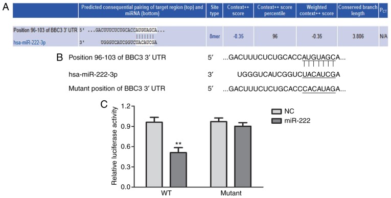
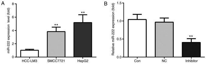
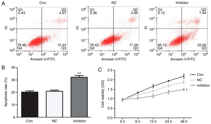
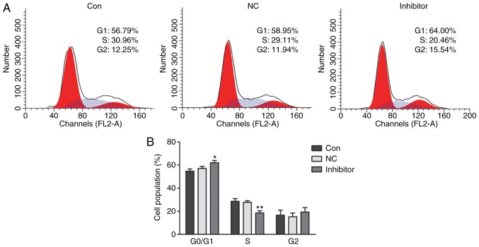
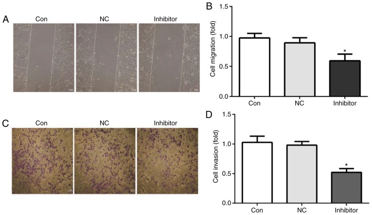
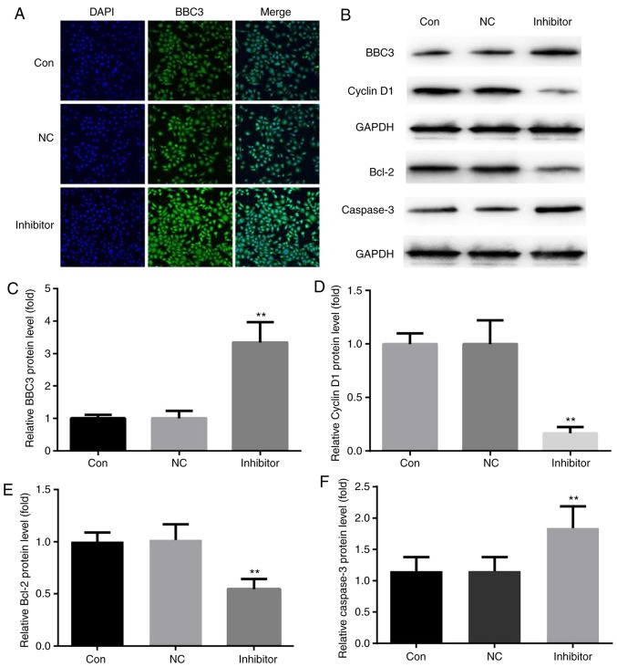
The present study aimed to investigate molecular mechanisms associated with liver cancer and provide a possible therapeutic target for the treatment of liver cancer. Liver cancer patients that were diagnosed and treated at the Central Hospital of China National Petroleum Corp. were included in the present study. microRNA (miR)‑222 was predicted to target B‑cell lymphoma-2 (Bcl‑2) binding component 3 (BBC3, also known as p53 upregulated modulator of apoptosis) by a bioinformatics analysis with TargetScan, which was verified by a dual‑luciferase reporter assay system. The correlations between BBC3 and miR‑222 levels and the patients' characteristics were analyzed. Furthermore, reverse transcription‑quantitative polymerase chain reaction was used to assess the mRNA levels of miRNA‑222 in the HCC‑LM3, MHCC97H and HepG2 cell lines. HepG2 cells were then transfected with miR‑222 inhibitor or miR‑negative control inhibitor. Cell proliferation, apoptosis, cell cycle, migration and invasion were evaluated by an MTT assay, flow cytometry, wound healing assay and Transwell assay, respectively. BBC3 was quantified by immunofluorescence and western blot analysis, and cyclin D1, Bcl‑2 and caspase‑3 levels were also evaluated by western blotting. miR‑222 inhibitor obviously inhibited HepG2 cell proliferation, migration, invasion, BBC3 and cyclin D1 protein expression levels and enhanced HepG2 cell apoptosis as well as the protein levels of Bcl‑2 and caspase‑3. miR‑222 level in tumors ≥5 cm (maximum) was significantly higher compared with tumors <5 cm (maximum) and was significantly higher in metastatic tumors compared with non‑metastatic tumors, while BBC3 level showed the adverse changes. The results of the present study suggested that miR‑222 inhibitor exerted anti‑cancer effects against liver cancer cells, probably by targeting the 3' untranslated region (UTR) of BBC3.


Figures

Figure 1
Figure 1
miR-222 targets the 3′UTR of the mRNA of BBC3. (A) miR-222 was predicted to target the mRNA of BBC3 3′UTR. (B) The mutation introduced in the 3′UTR of the mRNA of BBC3. (C) The relative luciferase activity in HepG2 cells that were transfected with miR-222 inhibitor and pmir-BBC3wt-3′UTR was obviously lower than that in HepG2 cells which were transfected with miR-222 inhibitor and pmir-BBC3mut-3′UTR. **P<0.01 miR-222 group vs. NC group. miR/miRNA, microRNA; BBC3, B-cell lymphoma 2 binding component 3; UTR, untranslated region; mut, mutant; wt, wild-type; NC, negative control; hsa, Homo sapiens.
Figure 2
Figure 2
miR-222 expression is highest in the HepG2 cell line. (A) Among 3 human liver cancer cell lines, including HCC-LM3, SMCC7721 and HepG2, the latter had the highest level of miR-222. Therefore, HepG2 cells were used in the subsequent experiments. (B) No significant difference in the expression levels of miR-222 was detected between the control group and miR-NC inhibitor group, but it was notably decreased in the miR-222 inhibitor group. **P<0.01 miR-222 inhibitor group vs. NC group. miR, microRNA; NC, negative control.
Figure 3
Figure 3
miR-222 inhibitor increases the apoptotic rate of HepG2 cells and decreases their proliferation. No obvious changes in HepG2 cell apoptosis or proliferation were identified between the control and the miR-NC inhibitor group. (A and B) Compared with HepG2 cells in the control group and miR-NC inhibitor group, the apoptotic rate was significantly increased by treatment with miR-222 inhibitor, while (C) the proliferation ability was significantly reduced by miR-222 inhibitor. **P<0.01 miR-222 inhibitor group vs. NC group. miR, microRNA; NC, negative control; PI, propidium iodide; Con, control; FITC, fluorescein isothiocyanate; OD, optical density; Q, quadrant.
Figure 4
Figure 4
miR-222 inhibitor induces cell cycle arrest of HepG2 cells at G0/G1 phase. (A) Cell cycle distribution profiles and (B) quantified populations in each phase of the cell cycle are presented. No obvious changes in the cell populations in each phase were observed between the control group and the miR-NC inhibitor group. Furthermore, no notable difference in the number of cells in G2 phase was observed between the 3 groups. However, the G0/G1 phase population in the miR-222 inhibitor group (64.00%) was significantly increased compared with that in the control (56.79%) and miR-NC group (58.95%); and the S-phase population in the miR-222 inhibitor group (20.46%) was decreased in comparison with that in the control (30.96%) and miR-NC group (29.11%). *P<0.05, **P<0.01 miR-222 inhibitor group vs. NC group. miR, microRNA; NC, negative control.
Figure 5
Figure 5
miR-222 inhibitor impairs HepG2 cell migration and invasion. (A and B) The cell migration ability was tested by a wound healing assay and (C and D) the cell invasion ability of was assessed using Transwell chambers. The results indicated that, compared with HepG2 cells in the control and miR-NC inhibitor group, the cell migration and invasion ability were significantly reduced by miR-222 inhibitor (scale bar, 100 µm). miR, microRNA; NC, negative control; Con, control. *P<0.05 miR-222 inhibitor group vs. NC group. miR, microRNA; NC, negative control.
Figure 6
Figure 6
miR-222 inhibitor promotes the expression of BBC3 in HepG2 cells. (A) Compared with that in HepG2 cells in the control and miR-NC inhibitor groups, miR-222 inhibitor significantly increased the immunofluorescence intensity of BBC3 (magnification, ×400). (B) Western blot analysis indicated that compared with HepG2 cells in the control and miR-NC inhibitor groups, miR-222 inhibitor decreased Bcl-2 and cyclin D1 protein levels, increased the protein levels of BBC3 and cleaved caspase-3 in HepG2 cells. (C–F) The corresponding statistical data of BBC3, cyclin D1, Bcl-2, and cleaved caspase-3 were exhibited. Bcl-2, B-cell lymphoma 2; miR, microRNA; NC, negative control; Con, control; BBC3, Bcl-2 binding component 3. **P<0.01 miR222 inhibitor group vs. NC group.

References

    1. Adult Primary Liver Cancer Treatment (PDQ®)-Patient Version. NCI. 6 July 2016. Archived from the original on October 2016. Retrieved 29 September 2016.
    1. World Cancer Report 9283204298World Health Organization. 2014;2014(6):5.
    1. Tsujimoto Y, Finger LR, Yunis J, Nowell PC, Croce CM. Cloning of the chromosome breakpoint of neoplastic B cells with the t (14;18) chromosome translocation. Science. 1984;226:1097–1099. doi: 10.1126/science.6093263. - DOI - PubMed
    1. Cleary ML, Smith SD, Sklar J. Cloning and structural analysis of cDNAs for bcl-2 and a hybrid bcl-2/immunoglobulin transcript resulting from the t (14;18) translocation. Cell. 1986;47:19–28. doi: 10.1016/0092-8674(86)90362-4. - DOI - PubMed
    1. Yu J, Zhang L, Hwang PM, Kinzler KW, Vogelstein B. PUMA induces the rapid apoptosis of colorectal cancer cells. Mol Cell. 2001;7:673–682. doi: 10.1016/S1097-2765(01)00213-1. - DOI - PubMed

MeSH terms