Review
. 2018 Jul;9(4):e1476.
doi: 10.1002/wrna.1476. Epub 2018 Apr 25.

Alternative-splicing defects in cancer: Splicing regulators and their downstream targets, guiding the way to novel cancer therapeutics

Affiliations
Review

Alternative-splicing defects in cancer: Splicing regulators and their downstream targets, guiding the way to novel cancer therapeutics

Laura M Urbanski et al. Wiley Interdiscip Rev RNA. 2018 Jul.

Abstract

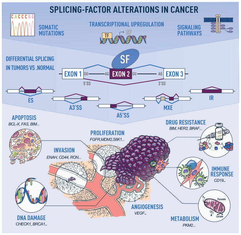
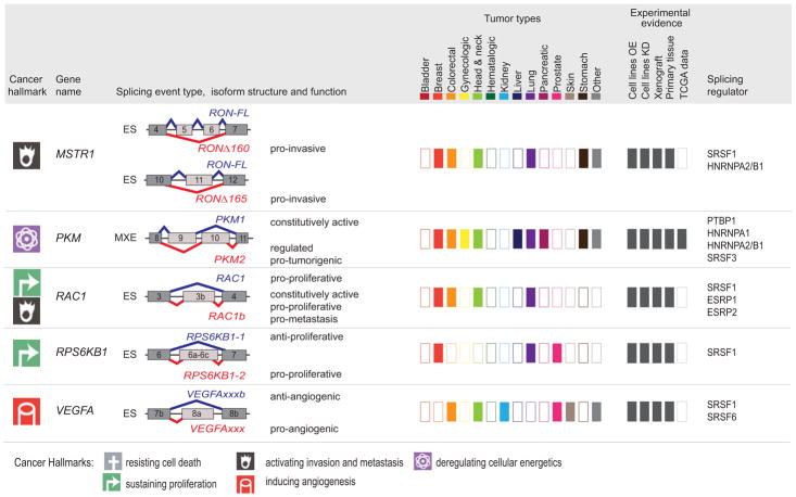
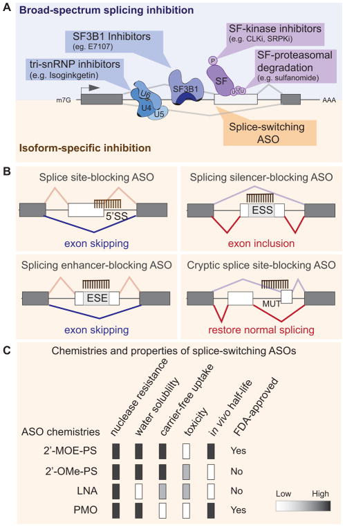
Defects in alternative splicing are frequently found in human tumors and result either from mutations in splicing-regulatory elements of specific cancer genes or from changes in the regulatory splicing machinery. RNA splicing regulators have emerged as a new class of oncoproteins and tumor suppressors, and contribute to disease progression by modulating RNA isoforms involved in the hallmark cancer pathways. Thus, dysregulation of alternative RNA splicing is fundamental to cancer and provides a potentially rich source of novel therapeutic targets. Here, we review the alterations in splicing regulatory factors detected in human tumors, as well as the resulting alternatively spliced isoforms that impact cancer hallmarks, and discuss how they contribute to disease pathogenesis. RNA splicing is a highly regulated process and, as such, the regulators are themselves tightly regulated. Differential transcriptional and posttranscriptional regulation of splicing factors modulates their levels and activities in tumor cells. Furthermore, the composition of the tumor microenvironment can also influence which isoforms are expressed in a given cell type and impact drug responses. Finally, we summarize current efforts in targeting alternative splicing, including global splicing inhibition using small molecules blocking the spliceosome or splicing-factor-modifying enzymes, as well as splice-switching RNA-based therapeutics to modulate cancer-specific splicing isoforms. This article is categorized under: RNA in Disease and Development > RNA in Disease RNA Processing > Splicing Regulation/Alternative Splicing.

Keywords: RNA binding proteins; RNA biology; RNA-based therapies; alternative splicing; antisense oligonucleotides; cancer; isoforms; oncogenes; spliceosome; splicing factors; tumor suppressors.


Figures

Figure 1
Figure 1. Alternative-splicing alterations in cancer
Human tumors exhibit recurrent mutations in, or changes in the levels of, splicing regulatory factors, the latter of which can occur due to copy number changes, or alterations in the transcriptional, post-transcriptional, or post-translational regulation of splicing factors in response to signaling changes (top panel). These changes in splicing-factor levels lead to alterations in the splicing of their downstream targets, promoting events that follow one of the following patterns: exon skipping (ES), alternative 5′ or 3′ splice site (SS) selection (A5′SS or A3′SS), inclusion of mutually exclusive exons (MXE), or intron retention (IR) (middle panel). Misregulated splicing of isoforms involved in key cellular pathways contributes to tumor initiation and progression. Examples of cancer hallmarks and associated tumor isoforms are indicated (bottom panel).
Figure 2
Figure 2. Components of the core and regulatory splicing machinery that exhibit alterations in human tumors
(A) Graphical representation of the stepwise assembly of spliceosomal complexes on a pre-mRNA molecule and catalysis of the splicing reaction to generate mature spliced mRNA. First, the ATP-independent binding of U1 snRNP to the 5′ splice site (5′SS) of the intron initiate the assembly of the “Early” or E complex on the pre-mRNA. In addition, SF1 and U2AF2 bind respectively to the branch point site (BPS) and the polypyrimidine tract (Py-tract). In the second step, the ATP-dependent interaction of U2 snRNP with the BPS leads to the formation of the A complex. This interaction is stabilized by the SF3a and SF3b protein complexes, as well as U2AF2 and U2AF1, and leads the displacement of SF1 from the BPS. Recruitment of the pre-assembled U4/U6/U5 tri-snRNP marks the formation of the catalytically inactive B complex. Major conformational changes, including release of U1 and U4, lead to spliceosome activation and formation of the B* complex. The first catalytic step of splicing, generates the C complex and results in the formation of the lariat. Complex C performs the second catalytic step of splicing, which results in the joining of the two exons. Post-splicing the spliceosome disassembles in an orderly manner, releasing the mRNA, as well as the lariat bound by U2/U5/U6. The snRNP are then further dissociated and recycled. (B) Spliceosomal core factors that exhibit recurrent somatic mutations in human tumors are listed next each complex (colored boxes) and are shown in more details for complexes E and A (right panels). In addition to core splicing factors, regulatory splicing factors (SF) that can bind to exonic or intronic splicing enhancer (ESE or ISE) or silencer (ESS or ISS) sequences to fine-tune splicing are also found altered in human tumors (grey boxes).
Figure 3
Figure 3. Recurrent splicing-factor alterations detected in human tumors
Genomic alterations including expression changes and recurrent somatic mutations in splicing factors detected in more than 2% of tumors in several cohorts of patients, including TCGA data, are indicated per tumor type. Splicing-factor upregulation are depicted in red, downregulation in blue, and somatic mutations in green (See legend for details). Several splicing factors can be found both upregulated and downregulated in tumors of the same tissue, suggesting that distinct splicing-factor genomic alterations are associated with distinct tumor subtypes within the same tissue. AML: acute myeloid leukemia; AML/MDS: acute myeloid leukemia myelodysplastic syndrome; CMML: chronic myelomonocytic leukemia; HN: head and neck; MDS w/o RS: myelodysplastic syndrome without ringed sideroblasts; RARS/RCMD: refractory anemia with ringed sideroblasts and refractory cytopenia with multilineage dysplasia and ringed sideroblasts; MPN: myeloproliferative neoplasm. See references in text.
Figure 4
Figure 4. Defects in splicing-factor regulation lead to changes in splicing-factor levels, activity, and cellular localization
Schematic representation of the transcriptional, post-transcriptional, and post-translational steps that impact the expression of a splicing factor (SF). See text for specific examples and references.
Figure 5
Figure 5. Tumor-associated isoforms representative of the cancer hallmarks
Splicing event type, isoform structure, tumor expression, and experimental evidence for selected alternative splicing isoforms detected in human tumors. For simplicity, only the alternatively spliced sequences and the flanking exons and are shown (not at scale). The type of splicing event is indicated: ES: exon skipping; MXE: mutually exclusive exons; 5′ASS: 5′ alternative splice site selection; IR: intron retention. The corresponding isoforms are shown in red or blue and their respective functions are indicated when known, to the right of the schematic figure of the isoform. The cancer hallmark associated with the red isoform is indicated in the left-hand column (See legend for details). ‘Tumor types,’ indicates, using dark-colored rectangles, the expression of tumor-associated isoforms in each of the indicated tumor types (‘other tumors’ include: adrenal, gallbladder, ampullary, bone, and brain; ‘gynecological tumors’ include: ovarian, cervical, and uterine; ‘head and neck’ tumors include: oral, head and neck, tongue, esophageal, and thyroid). ‘Experimental evidence’ indicates, using dark gray rectangles, the expression and functional evidence for each isoform based the following experiments: (i) overexpression (OE) or knockdown (KD) in cell lines, (ii) tumor xenografts, (iii) expression in primary tissue, or (iv) expression in TCGA RNA-sequencing data. Known splicing regulatory proteins are listed for each gene. See text for references.
Figure 6
Figure 6. Therapeutic strategies to target splicing alterations in tumors rely either on broad-spectrum splicing inhibition or on isoform-specific modulation
(A) Small molecules targeting components of the spliceosome (e.g., SF3B1, or tri-sRNP) block their activity by preventing assembly of a functional spliceosome into the pre-mRNA, and thus globally inhibit splicing. Alternatively, broad splicing inhibition can be achieved by targeting the enzymes that modulating the activity of splicing regulatory factors (SF), using for example small molecules inhibitors of CLKs or SRPKs, two kinase families that regulate the phosphorylation and thus the activity of SR proteins. Compounds that affect splicing factor poly-ubiquitination and proteasomal degradation (e.g. sulfonamides) can also induce broad changes in splicing profiles. On the other hand, isoform specific inhibition can be achieved by using splice-switching antisense oligonucleotides (ASOs) that bind in a sequence-specific manner and modulate the outcome of a specific splicing isoform. (B) ASOs can promote exon skipping or inclusion by blocking the 5′SS, an exonic silencer (ESS), or enhancer element (ESE) or by preventing the usage of a mutant (MUT)/cryptic splice site. See text for details. (C) Properties of ASO chemistries are currently used for splicing-modulation. See text for details. 2′-MOE/PS: 2′-O-(2-methoxyethyl)/phosphorothioate; 2′-OMe/PS: 2′ O-methyl/phosphorothioate; PMO: phosphorodiamidate morpholino oligomer; LNA: locked nucleic acid.

References

    1. Biamonti G, Catillo M, Pignataro D, Montecucco A, Ghigna C. The alternative splicing side of cancer. Semin Cell Dev Biol. 2014;32:30–36. - PubMed
    1. Bertram K, et al. Cryo-EM Structure of a Pre-catalytic Human Spliceosome Primed for Activation. Cell. 2017;170(4):701–713 e711. - PubMed
    1. Hegele A, et al. Dynamic protein-protein interaction wiring of the human spliceosome. Mol Cell. 2012;45(4):567–580. - PubMed
    1. Zhang X, Yan C, Hang J, Finci LI, Lei J, Shi Y. An Atomic Structure of the Human Spliceosome. Cell. 2017;169(5):918–929 e914. - PubMed
    1. Wahl MC, Luhrmann R. SnapShot: Spliceosome Dynamics I. Cell. 2015;161(6):1474–e1471. - PubMed

Publication types

MeSH terms

Substances