. 2018 Aug 1;315(2):F364-F373.
doi: 10.1152/ajprenal.00063.2018. Epub 2018 Apr 25.

A translational model of chronic kidney disease in swine

Affiliations

A translational model of chronic kidney disease in swine

Alejandro R Chade et al. Am J Physiol Renal Physiol. .

Abstract

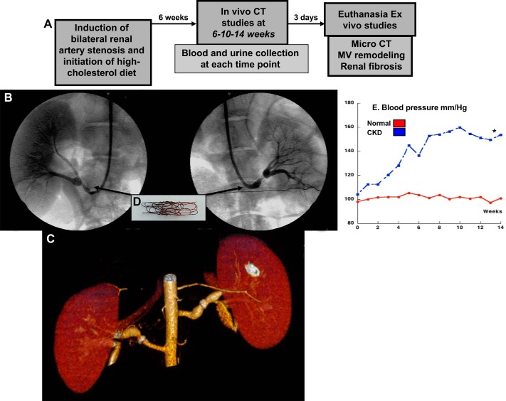
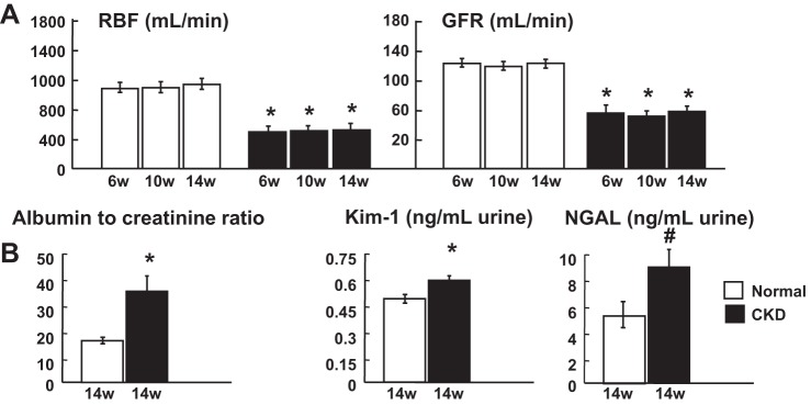
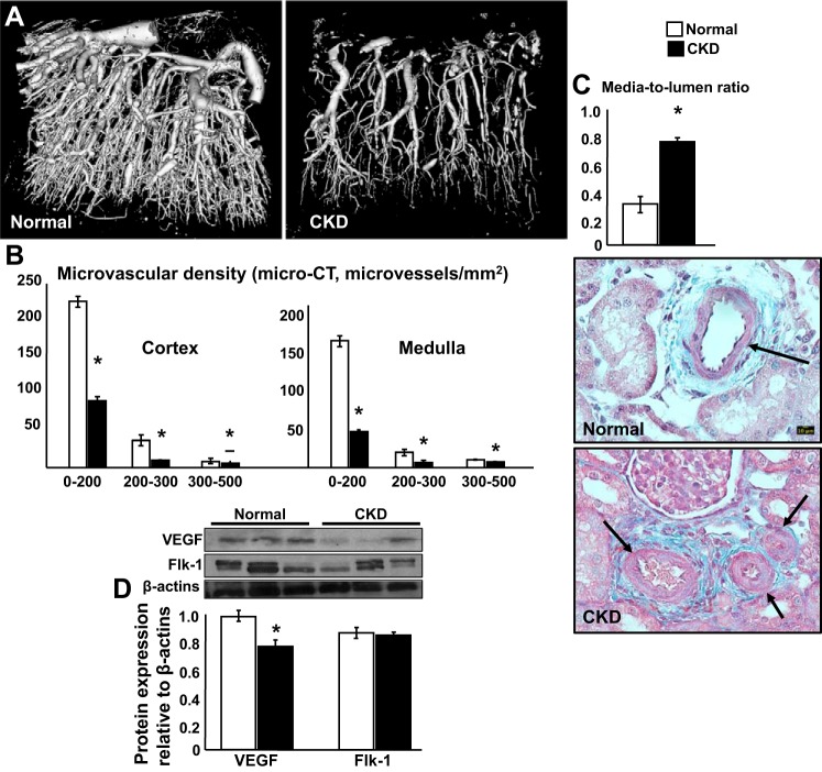
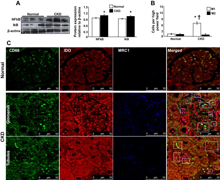
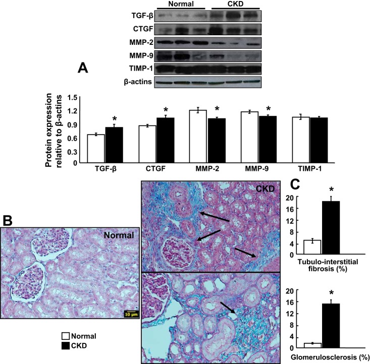
Animal models of chronic kidney disease (CKD) are critical for understanding its pathophysiology and for therapeutic development. The cardiovascular and renal anatomy and physiology of the pig are virtually identical to humans. This study aimed to develop a novel translational model of CKD that mimics the pathological features of CKD in humans. CKD was induced in seven domestic pigs by bilateral renal artery stenosis and diet-induced dyslipidemia. Animals were observed for a total of 14 wk. Renal hemodynamics and function were quantified in vivo using multi-detector CT after 6, 10, and 14 wk of CKD. Urine and blood were collected at each time-point, and blood pressure was continuously measured (telemetry). After completion of in vivo studies, pigs were euthanized, kidneys were removed, and microvascular (MV) architecture (μCT), markers of renal injury, inflammation, and fibrosis were evaluated ex vivo. Additional pigs were used as controls ( n = 7). Renal blood flow and glomerular filtration were reduced by 50% in CKD, accompanied by hypertension and elevated plasma creatinine, albumin-to-creatinine ratio and increased urinary KIM-1 and NGAL, suggesting renal injury. Furthermore, 14 wk of CKD resulted in cortical and medullary MV remodeling and loss, inflammation, glomerulosclerosis, tubular atrophy, and tubule-interstitial fibrosis compared with controls. The current study characterizes a novel model of CKD that mimics several of the pathological features observed in human CKD, irrespective of the etiology. Current approaches only slow rather than halt CKD progression, and this novel model may offer a suitable platform for the development of new treatments in a translational fashion.

Keywords: chronic renal disease; fibrosis; inflammation; microcirculation; renal hemodynamics.


Conflict of interest statement

No conflicts of interest, financial or otherwise, are declared by the authors.

Figures

Fig. 1.
Fig. 1.
A: design of the study. B and C: conventional renal angiography (arm; B and C) and CT angiography [multidetector computer tomography (MDCT); C] after 14 wk of chronic kidney disease (CKD). D and E: copper coil deployed inside each main renal artery on day 0 to induce renal artery stenosis (D), which resulted in a significant increase in blood pressure (measured by telemetry; E). *P < 0.05 vs. normal.
Fig. 2.
Fig. 2.
Experimental chronic kidney disease (CKD) resulted in a significant reduction of bilateral renal hemodynamics and function (compatible with CKD stage 2) and development of renal injury. A: renal blood flow (RBF) and glomerular filtration rate (GFR) after 6, 10, and 14 wk of CKD. B: albumin-to-creatinine ratio, urinary kidney injury molecule-1 (KIM-1), and neutrophil gelatinase associated lipocalin (NGAL) after 14 wk of CKD. *P < 0.05 vs. normal; #P = 0.07 vs. normal.
Fig. 3.
Fig. 3.
Experimental chronic kidney disease (CKD) induced significant renal microvascular rarefaction and remodeling in both kidneys. A and B: representative pictures of renal microvascular (MV) density (3-dimensional μCT reconstruction) and quantification (MV density of microvessels of diameter between 0 and 200, 200 and 300, and 300 and 500 μm. C: quantification of MV media to lumen ratio (both kidneys). D: representative protein expression (3 bands/group) of VEGF and Flk-1 receptor in normal pigs and pigs after 14 wk of CKD. *P < 0.05 vs. normal.
Fig. 4.
Fig. 4.
Experimental kidney disease (CKD) resulted in substantial inflammation in both kidneys. A: representative protein expression (3 bands/group) of proinflammatory NF-κB and IκB in normal pigs and pigs after 14 wk of CKD. B and C: representative quantification and pictures of renal sections costained with CD68 (total macrophages), indolamine 2,3-dioxygenase (IDO; M1 macrophages), manose receptor C type 1 (MRC1; M2 macrophages), and merged CD68/IDO/MRC-1 (×63) shown as examples to illustrate renal inflammatory infiltrates and identify M1 and M2 macrophages after 14 wk of CKD. The renal expression of NF-κB/IκB was increased in CKD compared with controls, whereas the presence of proinflammatory M1 macrophages (white boxes) was significantly higher than M2 (magenta boxes) in pigs after 14 wk of CKD. *P < 0.05 vs. normal; †P < 0.01 vs. M2.
Fig. 5.
Fig. 5.
Experimental kidney disease (CKD) induced significant glomerulosclerosis and tubule/interstitial fibrosis in both kidneys. A: representative protein expression (3 bands/group) of profibrotic transforming growth factor-β (TGFβ), connective tissue growth factor (CTGF), and tissue remodeling factors matrix metalloproteinase (MMP)-2, MMP-9, and tissue inhibitors of metalloproteinases (TIMP-1). Representative renal cross-sections stained with trichrome (×20), shown as examples to illustrate (B) and quantify (C) the degree of renal fibrosis after 14 wk of CKD. *P < 0.05 vs. normal.

References

    1. Abbott A. Pig geneticists go the whole hog. Nature 491: 315–316, 2012. doi:10.1038/491315a. - DOI - PubMed
    1. Akchurin OM, Kaskel F. Update on inflammation in chronic kidney disease. Blood Purif 39: 84–92, 2015. doi:10.1159/000368940. - DOI - PubMed
    1. Bábíčková J, Klinkhammer BM, Buhl EM, Djudjaj S, Hoss M, Heymann F, Tacke F, Floege J, Becker JU, Boor P. Regardless of etiology, progressive renal disease causes ultrastructural and functional alterations of peritubular capillaries. Kidney Int 91: 70–85, 2017. doi:10.1016/j.kint.2016.07.038. - DOI - PubMed
    1. Becker GJ, Hewitson TD. Animal models of chronic kidney disease: useful but not perfect. Nephrol Dial Transplant 28: 2432–2438, 2013. doi:10.1093/ndt/gft071. - DOI - PubMed
    1. Chade AR, Best PJ, Rodriguez-Porcel M, Herrmann J, Zhu X, Sawamura T, Napoli C, Lerman A, Lerman LO. Endothelin-1 receptor blockade prevents renal injury in experimental hypercholesterolemia. Kidney Int 64: 962–969, 2003. doi:10.1046/j.1523-1755.2003.00170.x. - DOI - PubMed

Publication types

MeSH terms

LinkOut - more resources