

S1P₄ Regulates Passive Systemic Anaphylaxis in Mice but Is Dispensable for Canonical IgE-Mediated Responses in Mast Cells
- PMID: 29693558
- PMCID: PMC5983835
- DOI: 10.3390/ijms19051279
S1P₄ Regulates Passive Systemic Anaphylaxis in Mice but Is Dispensable for Canonical IgE-Mediated Responses in Mast Cells
Abstract
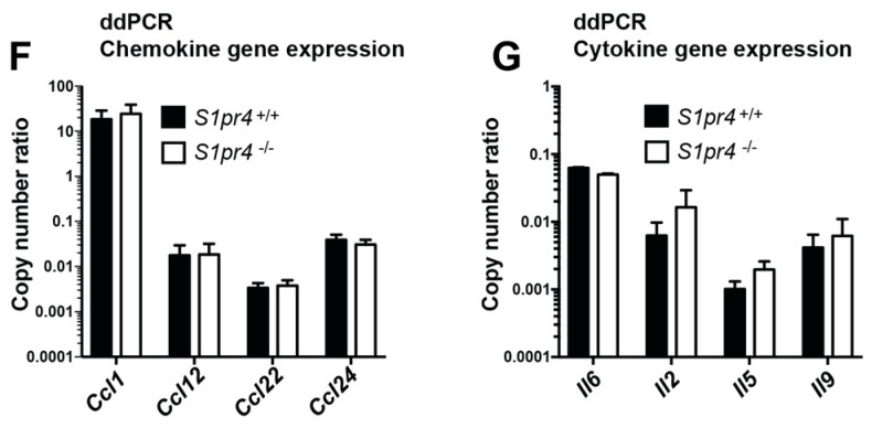
Mast cells are key players in the development of inflammatory allergic reactions. Cross-linking of the high-affinity receptor for IgE (FcεRI) on mast cells leads to the generation and secretion of the sphingolipid mediator, sphingosine-1-phosphate (S1P) which is able, in turn, to transactivate its receptors on mast cells. Previous reports have identified the expression of two of the five receptors for S1P on mast cells, S1P₁ and S1P₂, with functions in FcεRI-mediated chemotaxis and degranulation, respectively. Here, we show that cultured mouse mast cells also express abundant message for S1P₄. Genetic deletion of S1pr4 did not affect the differentiation of bone marrow progenitors into mast cells or the proliferation of mast cells in culture. A comprehensive characterization of IgE-mediated responses in S1P₄-deficient bone marrow-derived and peritoneal mouse mast cells indicated that this receptor is dispensable for mast cell degranulation, cytokine/chemokine production and FcεRI-mediated chemotaxis in vitro. However, interleukin-33 (IL-33)-mediated enhancement of IgE-induced degranulation was reduced in S1P₄-deficient peritoneal mast cells, revealing a potential negative regulatory role for S1P₄ in an IL-33-rich environment. Surprisingly, genetic deletion of S1pr4 resulted in exacerbation of passive systemic anaphylaxis to IgE/anti-IgE in mice, a phenotype likely related to mast cell-extrinsic influences, such as the high circulating levels of IgE in these mice which increases FcεRI expression and consequently the extent of the response to FcεRI engagement. Thus, we provide evidence that S1P₄ modulates anaphylaxis in an unexpected manner that does not involve regulation of mast cell responsiveness to IgE stimulation.
Keywords: IL-33; S1P4; S1pr4; anaphylaxis; chemotaxis; degranulation; mast cell; mediator release; sphingosine-1-phosphate.
Conflict of interest statement
The authors declare no conflict of interest.
Figures


Similar articles
-
Interrogation of sphingosine-1-phosphate receptor 2 function in vivo reveals a prominent role in the recovery from IgE and IgG-mediated anaphylaxis with minimal effect on its onset.Immunol Lett. 2013 Feb;150(1-2):89-96. doi: 10.1016/j.imlet.2013.01.005. Epub 2013 Jan 18. Immunol Lett. 2013. PMID: 23337656 Free PMC article.
-
Transgenic mice expressing the human high-affinity immunoglobulin (Ig) E receptor alpha chain respond to human IgE in mast cell degranulation and in allergic reactions.J Exp Med. 1996 Jan 1;183(1):49-56. doi: 10.1084/jem.183.1.49. J Exp Med. 1996. PMID: 8551243 Free PMC article.
-
Absence of platelet endothelial cell adhesion molecule-1 (CD31) leads to increased severity of local and systemic IgE-mediated anaphylaxis and modulation of mast cell activation.J Immunol. 2002 Jun 15;168(12):6455-62. doi: 10.4049/jimmunol.168.12.6455. J Immunol. 2002. PMID: 12055265
-
Unraveling the complexities of sphingosine-1-phosphate function: the mast cell model.Prostaglandins Other Lipid Mediat. 2008 Jun;86(1-4):1-11. doi: 10.1016/j.prostaglandins.2008.02.005. Epub 2008 Mar 4. Prostaglandins Other Lipid Mediat. 2008. PMID: 18403224 Free PMC article. Review.
-
A current understanding of Fc epsilon RI-dependent mast cell activation.Curr Allergy Asthma Rep. 2008 Mar;8(1):14-20. doi: 10.1007/s11882-008-0004-z. Curr Allergy Asthma Rep. 2008. PMID: 18377769 Review.
Cited by
-
A Critical Function for the Transcription Factors GLI1 and GLI2 in the Proliferation and Survival of Human Mast Cells.Front Immunol. 2022 Feb 16;13:841045. doi: 10.3389/fimmu.2022.841045. eCollection 2022. Front Immunol. 2022. PMID: 35251038 Free PMC article.
-
Aldh2 Attenuates Stem Cell Factor/Kit-Dependent Signaling and Activation in Mast Cells.Int J Mol Sci. 2019 Dec 10;20(24):6216. doi: 10.3390/ijms20246216. Int J Mol Sci. 2019. PMID: 31835486 Free PMC article.
-
Sphingolipids and their enigmatic role in asthma.Adv Biol Regul. 2018 Dec;70:74-81. doi: 10.1016/j.jbior.2018.09.001. Epub 2018 Sep 5. Adv Biol Regul. 2018. PMID: 30197277 Free PMC article. Review.
-
The Crosstalk between FcεRI and Sphingosine Signaling in Allergic Inflammation.Int J Mol Sci. 2022 Nov 11;23(22):13892. doi: 10.3390/ijms232213892. Int J Mol Sci. 2022. PMID: 36430378 Free PMC article. Review.
-
The Enigma of Sphingolipids in Health and Disease.Int J Mol Sci. 2018 Oct 12;19(10):3126. doi: 10.3390/ijms19103126. Int J Mol Sci. 2018. PMID: 30321983 Free PMC article. No abstract available.
References
-
- Allende M.L., Bektas M., Lee B.G., Bonifacino E., Kang J., Tuymetova G., Chen W., Saba J.D., Proia R.L. Sphingosine-1-phosphate lyase deficiency produces a pro-inflammatory response while impairing neutrophil trafficking. J. Biol. Chem. 2011;286:7348–7358. doi: 10.1074/jbc.M110.171819. - DOI - PMC - PubMed
-
- Gorlino C.V., Ranocchia R.P., Harman M.F., Garcia I.A., Crespo M.I., Moron G., Maletto B.A., Pistoresi-Palencia M.C. Neutrophils exhibit differential requirements for homing molecules in their lymphatic and blood trafficking into draining lymph nodes. J. Immunol. 2014;193:1966–1974. doi: 10.4049/jimmunol.1301791. - DOI - PubMed
MeSH terms
Substances
LinkOut - more resources
Full Text Sources
Other Literature Sources
Medical
Molecular Biology Databases
