Review
. 2018 Apr 25;7(2):48.
doi: 10.3390/pathogens7020048.

Adenylate Cyclases of Trypanosoma brucei, Environmental Sensors and Controllers of Host Innate Immune Response

Affiliations
Review

Adenylate Cyclases of Trypanosoma brucei, Environmental Sensors and Controllers of Host Innate Immune Response

Didier Salmon. Pathogens. .

Abstract

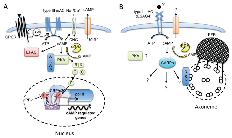
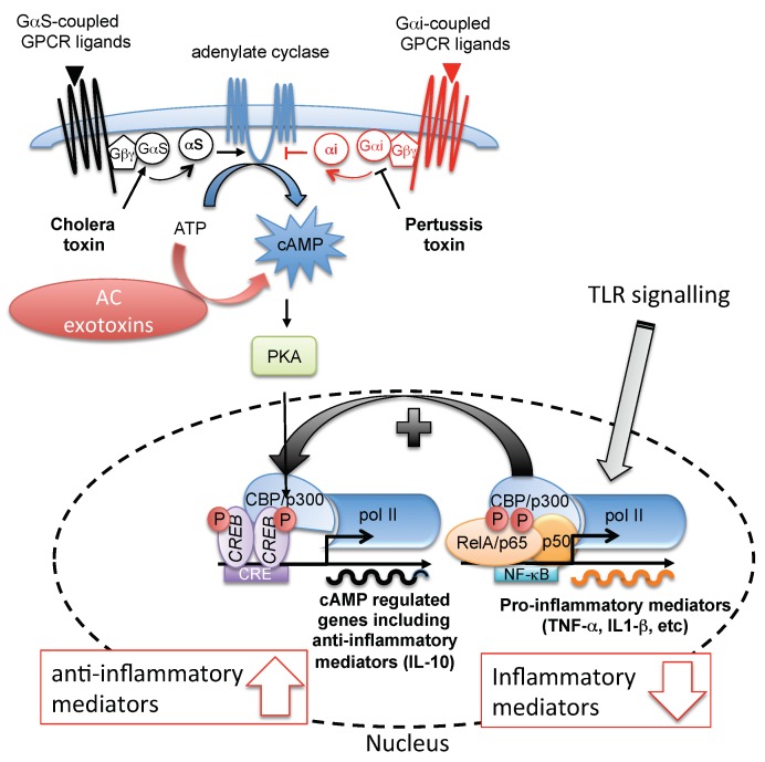
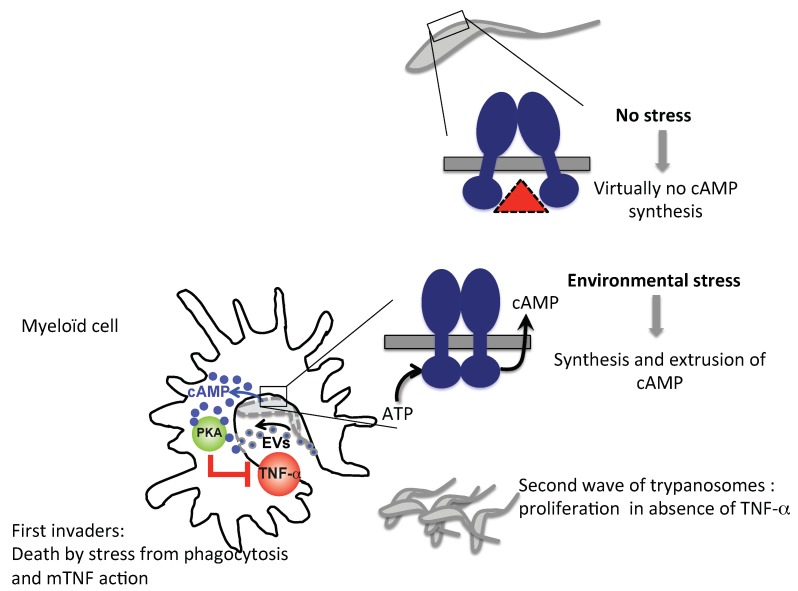
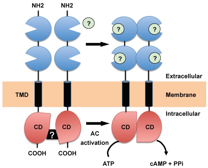
Trypanosoma brucei, etiological agent of Sleeping Sickness in Africa, is the prototype of African trypanosomes, protozoan extracellular flagellate parasites transmitted by saliva (Salivaria). In these parasites the molecular controls of the cell cycle and environmental sensing are elaborate and concentrated at the flagellum. Genomic analyses suggest that these parasites appear to differ considerably from the host in signaling mechanisms, with the exception of receptor-type adenylate cyclases (AC) that are topologically similar to receptor-type guanylate cyclase (GC) of higher eukaryotes but control a new class of cAMP targets of unknown function, the cAMP response proteins (CARPs), rather than the classical protein kinase A cAMP effector (PKA). T. brucei possesses a large polymorphic family of ACs, mainly associated with the flagellar membrane, and these are involved in inhibition of the innate immune response of the host prior to the massive release of immunomodulatory factors at the first peak of parasitemia. Recent evidence suggests that in T. brucei several insect-specific AC isoforms are involved in social motility, whereas only a few AC isoforms are involved in cytokinesis control of bloodstream forms, attesting that a complex signaling pathway is required for environmental sensing. In this review, after a general update on cAMP signaling pathway and the multiple roles of cAMP, I summarize the existing knowledge of the mechanisms by which pathogenic microorganisms modulate cAMP levels to escape immune defense.

Keywords: TNF-α; Trypanosoma brucei; adenylate cyclase; cAMP signaling; inflammation; innate immunity.


Conflict of interest statement

The author declares no conflict of interest.

Figures

Figure 1
Figure 1
Schematic representation of cAMP signaling in mammalian cells (A) vs. T. brucei (B), highlighting the contrast between canonical mammalian cAMP/PKA signaling pathway and the African trypanosomes’ cAMP signaling, which mainly concentrates in the flagellum and is characterized by the almost total absence of polII transcriptional regulation. (A) In mammals, ligand (triangle) binding activates GPCR, which undergoes a conformational change and then activates the G proteins by promoting the exchange of GDP/GTP associated with the Gα subunit, triggering its dissociation from the Gβ/Gγ dimer to activate type III AC. AC produces cAMP from ATP. High local levels of cytosolic cAMP lead to activation of PKA holoenzyme, which binds the AKAP through a hydrophobic dimerization domain of the PKA-R subunit, Epac or CNG channel. Upon cAMP binding to PKA-R, PKA-C subunits dissociate, then translocate to the cell nucleus, and induce the phosphorylation of transcription factors, such as CREB, to activate cAMP-driven genes. CREB inactivation is promoted by a phosphatase (e.g., PP-1). PDE and MRP decrease intracellular cAMP levels and counterbalance the intracellular cAMP effect. (B) In T. brucei, a putative ligand (circle) or membrane stress (hypotonic, acidic, proteolytic) activates flagellar type III AC (prototype ESAG4 in bloodstream form). This AC is topologically similar to receptor-type GC and produces cAMP from ATP. In T. brucei no classical PKA effector is activated by cAMP; instead, the cAMP targets are CARPs, components of unknown function, which participate in a putative novel cAMP signaling pathway. RSP represents an AKAP-like protein linked to the flagellar axoneme (RSP3/AKAP97-like); PFR represents the parafagellar rod structure of the flagellum, which is linked to PDEs (TbPDEB1/B2). No CNG channels or Epac have been characterized in trypanosomatids, and there is no evidence for cAMP secretion via membrane channels. AKAP, A-kinase anchoring protein; CBP, cAMP-binding protein; CARP, cAMP response protein; CNG, cyclic nucleotide-gated ion channel; CRE, cAMP-response elements; CREB, cAMP response element-binding protein; EPAC, exchange protein directly activated by cAMP; Gαs, stimulatory G protein alpha subunit; Gβγ, G protein beta gamma subunits; GPCR, G-protein-coupled receptor; MRP, multidrug resistance protein; PDE, phosphodiesterase; PFR, paraflagellar rod; PKA, protein kinase A; PolII, RNA polymerase II; RSP, radial spoke protein.
Figure 2
Figure 2
Pathogen strategies to counteract immunity by subversion of host cell cAMP signaling. Several extracellular bacterial pathogens possess virulence effectors, which increase cAMP levels in host cells, either by a G-protein modifying ADP ribosylation (pertussis toxin and cholera toxin) or by secreted AC exotoxins. Normally, the binding of an agonist (triangle) activates GPCR, which undergoes a conformational change leading to liberation of either the Gαs or Gαi subunit from the Gβγ subunit complex to, respectively, activate or inhibit the production of cAMP by AC. Pertussis toxin and cholera toxin produced by some pathogenic bacteria cause elevated cAMP levels through ADP-ribosylation of either the Gαi subunit to prevent AC inhibition or of the Gαs subunit to constitutively activate AC, respectively. TLRs binding of components of pathogenic bacteria (PAMPs) by TLRs triggers activation of NF-κB (RelA (p65)/p50) leading to transcriptional expression of pro-inflammatory mediators. Conversely, the production of high cellular cAMP levels by exotoxins increases the activation of CREB through PKA (driven anti-inflammatory mediator), which then competes with p65 for limiting amounts of CBP, resulting in fewer p65/CBP complexes, which are required for NF-κB activities that drive TNF-α expression (curved black arrow). CBP, cAMP-binding protein; CRE, cAMP-response elements; CREB, cAMP response element-binding protein; Gαi, inhibitory G protein alpha subunit; Gαs, stimulatory G protein alpha subunit; Gβγ, G protein beta gamma subunits; GPCR, G-protein-coupled receptor; NF-κB, nuclear factor-κB; PolII, RNA polymerase II.; RelA, Rel-associated protein; TLR, Toll-like receptor.
Figure 3
Figure 3
Proposed model to explain how the sacrifice of some parasites is thought to disable the innate immune response mediated by myeloid cells, allowing efficient host colonization by a second wave of invaders. In the absence of membrane stress, low basal levels of intracellular cAMP are produced following the combined actions of TbPDEB1/2 and low dimerization tendency of AC catalytic domains (red triangle). In the presence of stress, upon phagocytosis of the parasites by the acidic phagosome environment or following mTNF-mediated trypanolysis, CHDs dimerize, triggering a massive synthesis of cAMP that is translocated through an unknown mechanism into the myeloid cells cytosol or by phagocytosis/direct membrane fusion of extracellular vesicles (EVs) produced during phagocytic stress, blocking the synthesis of mTNF through activation of the host PKA. In this altruistic strategy, the sacrifice of the first pathogen invaders enables a second wave of trypanosomes to proliferate in the absence of local TNF-α, essential for the establishment of infection.
Figure 4
Figure 4
Predicted topology and hypothetical mode of activation of the receptor-type AC from T. brucei. A single transmembrane domain (TMD) separates an extracellular N-terminal domain containing two Venus flytrap (VFT) domains from a catalytic homology domain (CHD). AC activation is triggered by efficient head-to-tail dimerization of the CHDs that was postulated to be mediated through the N-terminal domains. This would involve conformational changes (e.g., dimerization) of VFT domains upon ligand binding (green circle) or membrane stress. Black box with question mark represents the allosteric inhibition mediated through the Δ-subdomain/phosphorylation. Membrane-bound forms appear to form homodimers and multimeric complexes [94].

Similar articles

Cited by

References

    1. Zhang Y., Agrebi R., Bellows L.E., Collet J.F., Kaever V., Grundling A. Evolutionary adaptation of the essential trna methyltransferase trmd to the signaling molecule 3′,5′-camp in bacteria. J. Biol. Chem. 2017;292:313–327. doi: 10.1074/jbc.M116.758896. - DOI - PMC - PubMed
    1. Gorke B., Stulke J. Carbon catabolite repression in bacteria: Many ways to make the most out of nutrients. Nat. Rev. Microbiol. 2008;6:613–624. doi: 10.1038/nrmicro1932. - DOI - PubMed
    1. Van Haastert P.J. Transduction of the chemotactic camp signal across the plasma membrane of dictyostelium cells. Experientia. 1995;51:1144–1154. doi: 10.1007/BF01944732. - DOI - PubMed
    1. Gancedo J.M. Biological roles of camp: Variations on a theme in the different kingdoms of life. Biol. Rev. Camb. Philos. Soc. 2013;88:645–668. doi: 10.1111/brv.12020. - DOI - PubMed
    1. Sutherland E.W., Jr., Wosilait W.D. Inactivation and activation of liver phosphorylase. Nature. 1955;175:169–170. doi: 10.1038/175169a0. - DOI - PubMed

LinkOut - more resources